Pterygium – definition, risk factors and treatment
For sutureless pterygium removal with Prof. Mimouni call 058-644-5151 at any hour
Main points:
- Pterygium is a degenerative disease of the outer eye surface associated with chronic exposure to ultraviolet light and especially common in a tropical climate.
- Bare-sclera excision of pterygium should no longer be performed because of the high recurrence rate associated with this procedure.
- Excision of pterygium with a conjunctival autograft is the accepted technique for treating pterygium with low recurrence rates and minimal complications.
- The most common complication of surgery to remove pterygium is recurrence of the pterygium, and this usually returns in the first year after the excision.
- Recurrence rates vary with different surgical techniques, and adjuvant treatments are used in an attempt to reduce recurrence rates.
- Caution is needed regarding the use of adjuvant treatments to reduce pterygium recurrence such as β radiation or mitomycin C because it is associated with long-term complications that can harm vision including scleral necrosis and endophthalmitis.
News and articles in the field of pterygium on the ophthalmologist site
Pterygium treatment
February 3, 2022Pterygium – treatment. When we talk about the growth of a pterygium, treating it takes on added importance. Pterygium is scar tissue on the surface of the conjunctiva that grows over the front part of
Pinguecula
April 1, 2017What is pinguecula? Pinguecula is a benign lesion of the conjunctiva of the eye that usually appears on the nasal and temporal sides (more common on the nasal side). What causes pinguecula in the eye? These lesions appear as a result of exposure to ultra
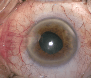
Pterygium surgery price
December 24, 2021How is a price set for pterygium surgery? Patients ask me what the price for pterygium surgery is? How is the cost of pterygium surgery determined? And how is a price set for pterygium surgery. The price is built according to several main factors:
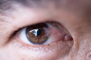
Natural treatment for pterygium
September 13, 2021Is there a natural treatment for pterygium? As a specialist in ophthalmology, with a subspecialty in diseases of the anterior segment of the eye, cornea and the outer surface of the eye, I often encounter patients with pterygium. They
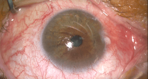
Pterygium removal
October 25, 2020Surgery to remove pterygium. The treatment of pterygium mainly includes surgery to remove the pterygium and approaches to reduce recurrence of the lesion after pterygium removal surgery. Various techniques, from the simplest procedure of bare-sclera excision
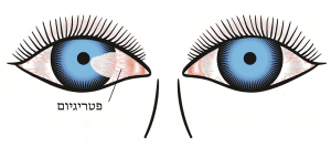
Pterygium excision
January 20, 2022Pterygium excision. Pterygium excision is a procedure in which pterygium is removed from the conjunctiva and cornea area. Pterygium is a non-cancerous growth of the conjunctiva of the eye, which involves the eye surface beyond the cornea.
Introduction to pterygium
Pterygium is a triangular "wing-like" growth composed of conjunctival epithelium and hypertrophic subconjunctival connective tissue that occurs on the nasal or temporal side in the area of the palpebral fissure and grows over the cornea. Pterygium remains an ocular enigma since the full pathogenic mechanisms remain unknown.
The symptoms of pterygium range from mild irritation and dryness to a decrease in vision as a result of irregular astigmatism or blurring of the visual axis. The difficulty in treating this deceptively benign disease stems from our lack of understanding of this condition and its tendency to recur after surgical excision. There is an abundance of surgical and medical means available in the treatment of pterygium.
For sutureless pterygium removal – contact Prof. Mimouni
 |  |
| Call 058-644-5151 at any hour | |
Definition and morphology
Pterygium can be defined as primary or recurrent. Recurrent pterygium is usually more aggressive and can occur several weeks to months after excision of a primary pterygium.
Pterygium consists of a "head" at its apex, usually with a cap of blood vessels at the leading edge. The "body" represents the main part of the pterygium over the sclera and extends from the canthus area. Pterygium usually appears on the nasal side, in the intra-palpebral fissure, but can also appear on the temporal side. A temporal pterygium can coexist alongside a nasal pterygium (a "double headed" pterygium). The subconjunctival or fibrovascular component of the pterygium may vary considerably in its appearance, from thin and transparent to thick and fleshy. A simple grading scale was developed based on slit-lamp examination and the relative transparency of the pterygium body that predicts recurrence. In this grading, T1 (atrophic) indicates a pterygium in which the episcleral vessels underlying the pterygium body are clearly visible.
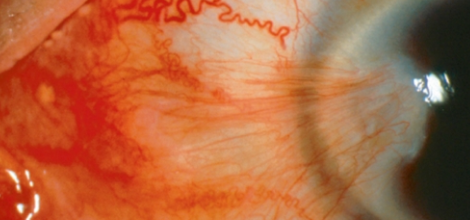
Grade T3 (fleshy) indicates a thick pterygium in which the episcleral vessel underlying the pterygium body is completely hidden by fibrovascular tissue.
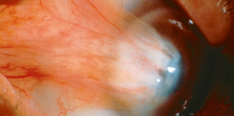
A pterygium in which the details of the episcleral vessel are seen unclearly or partially hidden is classified as grade T2 (intermediate).
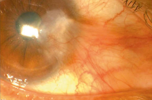
In a randomized clinical trial, bare-sclera excision was compared with a conjunctival autograft and they were able to show that pterygium recurrence is clearly related to the amount of fibrovascular tissue present in the pterygium when surgery was performed by the bare-sclera method. In the conjunctival autograft group there were too few recurrences to determine the significance in this form of procedure. A fleshy pterygium had a higher tendency to recur than a more transparent pterygium; the differences in recurrence rates were highly significant for both primary pterygium and recurrent pterygia.
Epidemiology of pterygium
Pterygium has a worldwide distribution but is more common in a hot and dry climate. Usually, pterygium occurs most often within the "pterygium belt" (latitude 37° north and south of the equator), where sun exposure significantly increases the risk of developing a pterygium. A meta-analysis of 20 studies that included 900 subjects in total demonstrated a pooled prevalence of pterygium of around 10% with an increased chance of pterygium in men (2.32 times for men) and with work in the sun (1.76 times for people who work frequently outdoors). It seems that the most common age of onset is in the twenties and thirties of life, and several studies showed that the prevalence of pterygium rises with age.
Risk factors
It seems that most of the risk factors are mainly environmental in nature, but there are studies that identified hereditary factors.
Ultraviolet radiation
The main environmental risk factor for the development of pterygium is exposure to ultraviolet light. This light, absorbed in the cornea and conjunctiva, promotes damage at the "cellular" level and leads to abnormal proliferation of cells.
Genetic factors
Reports of individual cases described clusters of family members with pterygium, and case-control studies showed that a family history is a risk factor, possibly even with an autosomal dominant inheritance pattern. However, the lack of a clear and consistent family connection indicates that pterygium does not have a strong familial basis. Recently, genes related to DNA repair, cell proliferation, migration and angiogenesis have been shown to be associated with pterygium. Not surprisingly, since disruption of cell repair and their proliferation may contribute to the multifactorial pathogenesis of pterygium.
Additional risk factors
Chronic irritation or inflammation occurring in the area of the limbus or the peripheral cornea has been proposed as a risk factor for pterygium. This theory suggests that chronic inflammation leads to a focal limbal stem cell deficiency and subsequent development of pterygium. There are also those who claim that there is the presence of an angiogenic factor that promotes the growth of pterygium. Recently, it has been demonstrated that vascular endothelial growth factor levels are higher in pterygium. There are studies that also showed a connection between the papilloma virus and pterygium.
Pathogenesis of pterygium
Is it a degenerative or proliferative disease?
Pterygium has long been considered a chronic degenerative condition, and this is based on histological examinations. This disease is classically described as "elastotic degeneration"; pterygium tissue is characterized by abnormal subepithelial tissue containing altered collagen fibers with positive staining for elastic stains. There are also clinical and etiological features of pterygium that point to a proliferative disorder, not very different from benign tumors, and a connection between pterygium and the p53 oncogene has been identified. It has also been shown that ultraviolet light, the main environmental risk factor in pterygium, causes mutations of the solar keratosis type, Bowen's disease and skin carcinomas. Furthermore, pterygium has a high tendency for aggressive recurrence after surgical excision, and adjuvant treatment with antimetabolites reduces its recurrence. It should be noted that the prevalence of conjunctival epithelial neoplasia in pterygium is about 1.8%.
Limbal stem cell deficiency (limbal stem cell deficiency) and pathological changes in the epithelium
The classic signs or clinical hallmarks of limbal stem cell deficiency include conjunctival growth over the cornea, growth of new blood vessels, chronic inflammation and destruction of the basement membrane. These signs are clearly present in pterygium, and therefore many researchers today have suggested that this is a manifestation of dysfunction or deficiency of local stem cells, perhaps as a result of destruction of stem cells associated with ultraviolet light.
Pterygium tissue demonstrates intrinsic pathologies of DNA repair as a result of ultraviolet light radiation, which is expressed in loss of heterozygosity of cells. Studies showed that basal epithelial layers of primary and recurrent pterygium display overexpression of the tumor-suppressor gene p53, an oncogene that acts as a transcription factor to activate or suppress the expression of genes controlling growth, thereby explaining the classic histological finding of epithelial hyperplasia seen in pterygium. It was also found that abnormal apoptosis occurs in the epithelium of primary and recurrent pterygium. Studies on pterygium also showed increased fibroblast activity with abnormal TGF-β signaling and overexpression of connective tissue metalloproteinases.
Treatment options for pterygium
There are several treatment options for pterygium, all of which are based on the principle of excision of the lesion combined with various methods to avoid recurrence of the pterygium.
The best-known treatments are bare-sclera excision, simple excision and closure of the conjunctiva or transposition of the conjunctiva, bare-sclera excision combined with anti-proliferative treatments, and excision of the lesion with reconstruction of the outer surface of the eye (for example pterygium excision combined with a conjunctival autograft, which is the accepted and most advanced treatment today).
On the page pterygium surgery one can read in great detail about treatment options. There is also the option to perform laser pterygium removal surgery.
Possible complications after pterygium removal surgery
Pterygium removal combined with a conjunctival autograft is usually considered a safe procedure, since serious vision-threatening complications usually do not develop. Early complications after surgery include graft edema, graft hemorrhages, graft retraction / suture breakage, graft inversion and graft necrosis. Conjunctival granulomas can also form a short time after surgery. Due to excessive inflammation and local irritation occurring at the site of exposed Tenon's tissue, granulomas may appear at the graft-harvest site, in the recipient bed adjacent to the conjunctival graft, or as a suture granuloma.
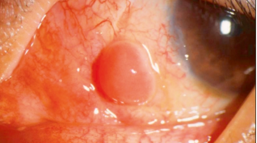
Late complications after surgery include epithelial inclusion cysts, scarring of the conjunctiva or fibrosis at the donor site, and elevated intraocular pressure caused by prolonged use of steroids as part of the post-surgery treatment.
Reasons for recurrence after conjunctival autograft
The morphological appearance of pterygium recurrence after conjunctival autograft transplantation indicates several factors that may lead to graft failure. Pterygium recurrence may occur at the upper or lower margins of the conjunctival graft, which suggests that pterygium tissue was excised insufficiently, or that the graft size was not sufficient. A thick graft including Tenon's tissue may lead to graft retraction and edge prolapse in the early post-surgery period, and cause exposure of bare sclera at the graft margins, which then becomes the site of recurrence. Early suture breakage will also lead to local graft retraction and local recurrence at this site. However, it should be noted that in certain cases a thin conjunctival graft that is aligned and sutured may subsequently become a recurrent pterygium in the limbus area, which indicates that some factor in the cornea or limbus may be responsible for recurrence in this case.
What is pterygium?

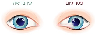
I am often asked what pterygium is. Pterygium (also known as surfer's eye) is a disease of the eye surface characterized mainly by a white or pinkish wing- or wedge-shaped growth of limbal tissue from the conjunctiva over the adjacent cornea. In most cases, pterygium grows from the inner corner of the eye (closest to the nose), but growth from the outer corner or from both sides of the eye simultaneously is sometimes possible. This is not malignant tissue and there is no invasion into the inner part of the eye or any other part of the body. The appearance of pterygium can range from a delicate transparent area with a few blood vessels, in the early stages, to a thick opaque growth that blocks vision in the later stages.
Pterygium consists of three separate parts: the cap, the head and the body / tail. The cap, or leading edge, is a flat area on the cornea composed mainly of fibroblasts that invade and destroy Bowman's membrane. The head is a vascular area located behind the cap and firmly attached to the cornea. The body/tail is the mobile area of the conjunctiva, which can be easily separated from the underlying tissue.
Histologically, the subepithelial tissue shows senile elastosis (basophilic degeneration) of the substantia propria with abnormal collagen fibers. There is a breakdown of Bowman's membrane, followed by invasion of the corneal surface. A unique feature of the epithelial cells of the pterygium is the evidence of the presence of various types of matrix metalloproteinases that are absent from the normal cells of the conjunctiva or cornea.
Usually the condition does not require any treatment unless it causes an actual disturbance in vision or constitutes severe discomfort. Even when no treatment is given, it is advisable to be under periodic monitoring by an ophthalmologist in order to make sure that the growth is not causing vision problems.
The exact cause of pterygium is still unknown. Many theories have been proposed about possible causes of pterygium, including the activation of special growth cells, called fibroblasts, by radiation, a deficiency of choline (which serves as a starting material for several important compounds in the cells of living organisms), an inflammatory disorder, a blood-vessel problem causing uncontrolled growth of blood vessels, immune-system disorders, disorders of the tear coating and even a possible viral connection.
Risk factors for pterygium
The risk factors for pterygium can be associated with the following conditions:
- Exposure to sunlight – the phenomenon is more common in people who spend a lot of time outdoors in hot weather, when the excessive exposure to ultraviolet radiation in sunlight is considered the cause.
- A dusty or sandy environment – long-term exposure to dry and wind-blown places is considered a factor that may contribute to the development and worsening of the condition.
- Age – the risk rises with age.
- Pterygium may be caused by a combination of several factors mentioned above.
What is pterygium – symptoms
In its early stages, pterygium is usually asymptomatic; however, when it is inflamed it may cause itching, a sensation as if there is a foreign body (rough) inside the eye, increased tearing, and burning. In later stages, the pterygium can grow over the iris and the pupil and cause blurring and disturbance in vision.
Diagnosis of pterygium
Pterygium can usually be diagnosed by its unique appearance and symptoms. However, in the early stages there is a possibility of confusion with similar growth phenomena affecting the conjunctiva such as pinguecula. If pterygium is diagnosed, a thorough eye examination should be performed to assess its effect on vision and to rule out less common diagnoses that can cause a growth in the eye.
What is pterygium – treatment?
The treatment of pterygium is initially limited to monitoring over time to see if it extends onto the visual axis. Symptoms of irritation, foreign-body sensation and tearing can be relieved with over-the-counter anti-allergy drops, lubricating eye drops, artificial tears, anti-inflammatory preparations, and ointment.

Surgery is the only way to remove a pterygium. If the eye drops and ointments do not provide sufficient relief, surgery may be recommended to remove the pterygium. Surgery is also recommended when the phenomenon causes impairment of vision. In such a case it is recommended to perform the surgery before the pterygium grows over the cornea, because then the surgery can scar the cornea and cause permanent vision problems. Sometimes the removal of the pterygium is also done for cosmetic reasons. During the surgery, the pterygium is carefully removed and a part of the conjunctiva is taken from under the eyelid and transplanted onto the area where the pterygium was. The surgery is performed under local anesthesia and lasts about 30 minutes.
In certain cases, the pterygium can return after it has been removed surgically, so surgical intervention should be considered seriously. Also, the eye may feel dry and irritated after the surgery. Medications can be prescribed to provide relief and reduce the risk of the pterygium growing back.
Prevention of pterygium
Where possible, one should avoid exposure to environmental factors that may cause pterygium by using sunglasses or a hat to protect the eyes from sunlight, wind and dust. If one wears glasses, they, like sunglasses, should provide protection against the sun's ultraviolet rays (UV). If a pterygium is already present, limiting exposure to sunlight, wind, dust, powder and smoke can slow its growth.
Prof. Michael Mimouni is an ophthalmologist specializing in pterygium with many years of experience in diagnosing, treating and operating on pterygium of the eye using the most advanced methods.
Have a question about your eyes? Want a specialist opinion?