A corneal transplant is a surgical procedure in which a damaged or diseased cornea is replaced with donated corneal tissue (the graft). When the whole cornea is replaced it is called penetrating keratoplasty (Penetrating Keratoplasty), and when only part of the cornea is replaced it is called lamellar keratoplasty (Lamellar Keratoplasty). Keratoplasty simply means surgery on the cornea. The graft is taken from a person who died recently without known eye diseases or other factors that could affect the survival chances of the donated tissue or the health of the recipient.
News and articles in the field of corneal transplantation
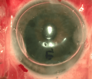
Corneal transplant DMEK
October 20, 2020What is DMEK? A corneal transplant of the DMEK type, short for Descemet Membrane Endothelial Keratoplasty, is a partial-thickness corneal transplant procedure that includes selective removal of the Descemet membrane and endothelium, followed by transplantation of corneal endothelium
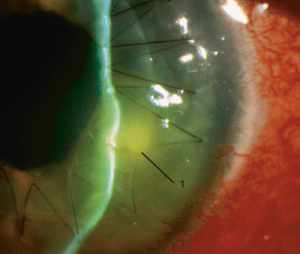
Corneal transplant risks
December 18, 2021There are various potential risks and complications for a corneal transplant depending on the type of surgery, the surgeon's experience and the complexity of the eye and the systemic background of the patient. In addition, in every type of surgery, there are complications that can occur during the surgery

Corneal transplant cost
February 10, 2022Is there really a cost for a corneal transplant? If the procedure is performed in a public hospital then the cost of a corneal transplant is nil (the cost of a corneal transplant in a public hospital: free for the patient).

Partial corneal transplant
December 6, 2021Surgical options for partial-thickness corneal transplant. In certain cases, it may not be necessary to perform a full-thickness corneal transplant surgery to treat a disease or damage in the cornea. Some patients can undergo a corneal transplant
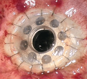
Artificial corneal transplant
December 4, 2021Background for artificial corneal transplant. An artificial corneal transplant is based on the use of a corneal graft that is made, at least in part, of a synthetic raw material that comes to provide an optical, structural and functional solution for patients who suffer
The cornea is the transparent front part of the eye covering the iris, the pupil and the anterior chamber. The surgical procedure is performed by ophthalmologists who specialize in corneal transplants and is often done on an inpatient basis in Israel, though in North America it is a procedure performed as day surgery. Donors can be of any age, though it is usually accepted to be at least two years of age, and there is no limit after that (Shimon Peres donated his corneas after he died). The corneal transplant is performed when non-invasive treatment such as medications or less-invasive surgeries (such as corneal cross-linking in keratoconus) cannot heal the cornea sufficiently.
Historical background
The first corneal transplant was performed in the year 1905 by Eduard Zirm (the Olomouc eye clinic, today the Czech Republic), and made it one of the first types of transplant surgeries of any organ that were performed successfully. The attempts of the Russian ophthalmologist Vladimir Filatov to transplant a cornea began with the first attempt in the year 1912 and continued, and gradually improved until on 6 May 1931 he successfully transplanted a cornea to a patient using corneal tissue from a person who had died. He reported extensively on another transplant in the year 1936, and shared his technique in full detail. In the year 1936, Castroviejo performed a first transplant in an advanced case of keratoconus, achieving a significant improvement in the patient's vision.
Tudor Thomas, a clinical teacher of the Welsh National School of Medicine, conceived the idea of a donor system for corneal transplants, and the East Grinstead bank was established in the year 1955. Advances in the operation of microscopes allowed surgeons to operate with a more magnified view, while advances in materials science allowed them to use sutures finer than a human hair.
What led to the success of corneal transplants was the establishment of eye banks. These are organizations located around the world to coordinate the distribution of donated corneas to surgeons, as well as to provide eyes for research. In Israel, as of 2020, there is no single central eye bank in the whole country, and most hospitals provide the corneas for themselves, while there are also sometimes collaborations between the various hospitals.
Indication for corneal transplant
General indications for a corneal transplant can be classified into 4 categories:
Optical: to improve visual acuity by replacing the unhealthy/abnormal tissue with healthy donor tissue. The most common indication in this category is pseudophakic bullous keratopathy (Pseudophakic bullous keratopathy), followed by keratoconus (keratoconus), Fuchs' dystrophy, and scarring due to keratitis and trauma.
Tectonic: to preserve the normal anatomy of the cornea and its integrity in patients with thinning of the stroma or after perforation of the cornea following a penetrating wound.
Therapeutic: to remove inflamed/infected corneal tissue that does not respond to treatment by conservative treatment such as antibiotics or anti-inflammatory treatment or antiviral treatment.
Cosmetic: to improve the appearance of patients with scars/opacities in the cornea (this is a minority of cases because of the risks in a corneal transplant and because of a lack of tissue).
Types of corneal transplant
Full-thickness corneal transplant: penetrating keratoplasty (Penetrating Keratoplasty). A surgery in which all the layers of the cornea are removed from a patient and all the layers from a donor's cornea are transplanted in their place. Today this type of corneal transplant is diminishing because there are today partial-thickness corneal transplants that have lower risks and, in the right indications, lead to better visual acuity results. Today the main indications for penetrating keratoplasty are: central full-thickness corneal scars as a result of trauma or infections, degenerative diseases of the cornea involving all the layers, severe chemical injuries of the cornea, repeat corneal transplant, and failure to perform an anterior lamellar corneal transplant such as DALK.
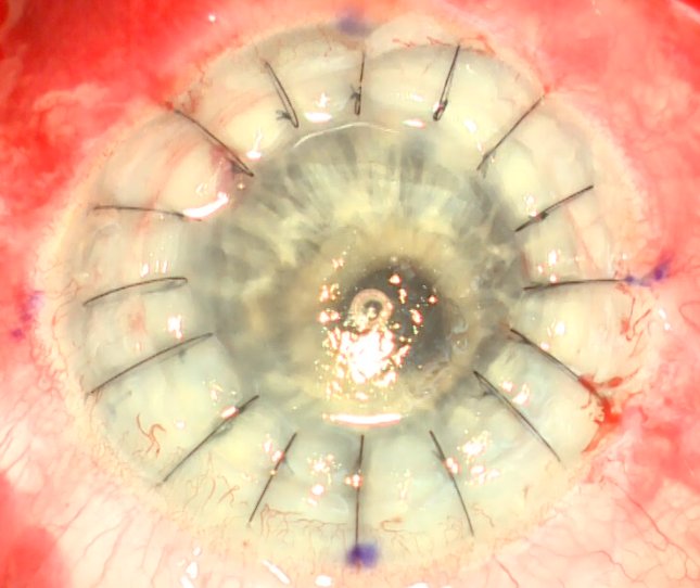
Partial corneal transplant: there are several types of common corneal transplants that include partial endothelial corneal transplants such as DMEK and DSAEK. There are also anterior partial corneal transplants such as DALK (more common and which we will elaborate on) and SALK (less common and which we will mention briefly).
Corneal transplant DMEK: this is the most advanced partial corneal transplant (as of the year 2020). The initials are short for Descemet Membrane Endothelial Keratoplasty. In this surgery the endothelium and Descemet membrane layer is removed from the patient and replaced with the same layers from a donor who died. The main indications for this surgery today are pseudophakic bullous keratopathy and Fuchs' dystrophy. This surgery has faster recovery of visual acuity, better final visual acuity and lower chances of graft rejection compared with DSAEK. However, it is a procedure that is more technically difficult to perform and is not suitable for complex eyes (long eyes, eyes after vitrectomy, eyes without a lens (aphakic) or with a large iris defect (aniridia)). In addition, there is a higher percentage of the need for Rebubbling (filling the anterior chamber with air) after the surgery compared with DSAEK.
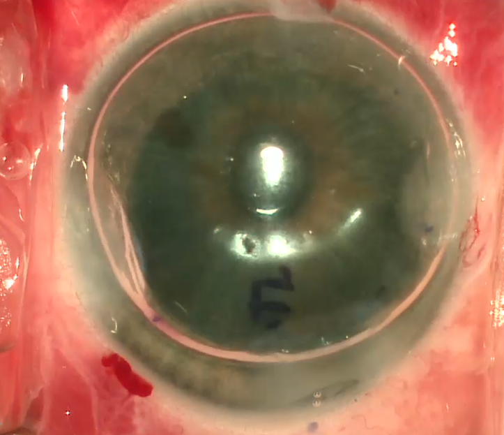
Corneal transplant DSAEK: this is a posterior partial corneal transplant, where the initials are short for Descemet Stripping Automated Endothelial Keratoplasty. In this surgery the endothelium layer and Descemet membrane are removed from the patient and replaced with endothelium, Descemet membrane and a little posterior stroma from the donor tissue. If the thickness of the stroma taken from the donor is below 100 microns then it is called Ultra thin DSAEK. If less than 60 microns then it is called nano thin DSAEK. The indications for this surgery are identical to those of DMEK, and when it is a very complex eye as mentioned above, it is preferable to perform DSAEK rather than DMEK. It is a procedure with lower chances of complications including graft rejection compared with penetrating keratoplasty.
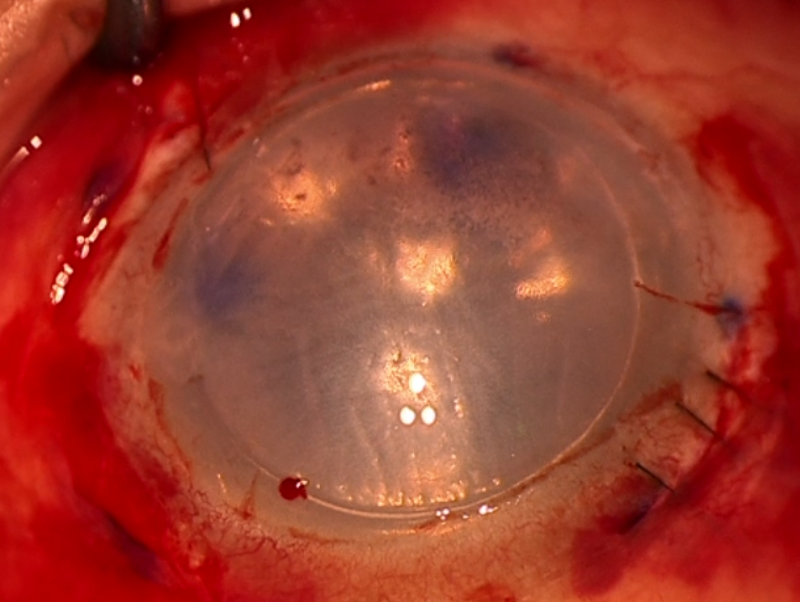
Corneal transplant DALK: this is an anterior partial corneal transplant, where the initials are short for Deep Anterior Lamellar Keratoplasty. In this surgery all the layers of the cornea are removed except for the endothelium, Descemet membrane and the Dua layer (the most posterior part of the stroma) and replaced with donor tissue from which the endothelium layer and Descemet membrane have been removed. The main indications for this surgery are keratoconus and corneal scars that are not full-thickness. It is a procedure with lower chances of complications involved in the Open Sky stage of penetrating keratoplasty and lower rejection chances as well.
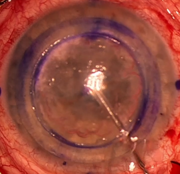
Corneal transplant SALK: this is an anterior corneal transplant, where the initials are short for Superficial Anterior Lamellar Keratoplasty. In this surgery only part of the anterior stroma is removed and replaced with similar tissue from a donor. The main indication is a very superficial scar. It is a procedure that is not performed much today.
Artificial corneal transplant
Boston Keratoprosthesis – Boston Keratoprosthesis
Boston KPro type 1 – posterior plate of titanium: the Boston keratoprosthesis is the most common synthetic cornea to date, with over 900 procedures performed around the world in the year 2008. The Boston KPro was developed at the eye and ear hospital under the leadership of Dr. Claes Dohlman.
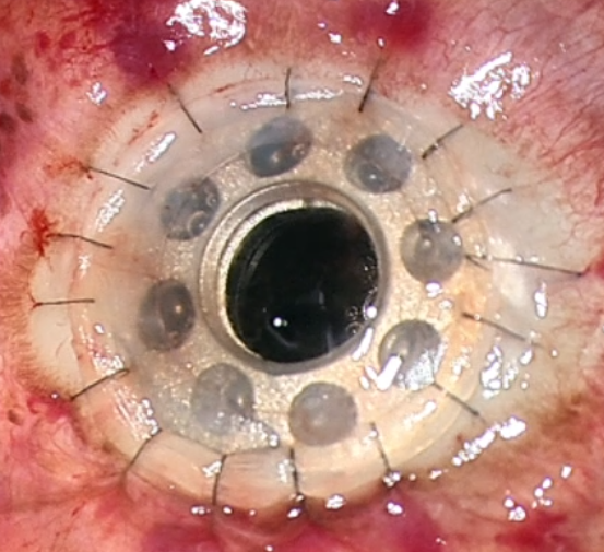
AlphaCor – AlphaCor: in cases where there were several graft failures or the risk for keratoplasty is high, synthetic corneas can successfully replace donor corneas. Such a device contains a peripheral skirt and a central transparent zone. These two parts are connected at the molecular level by an interpenetrating polymer network, made of poly(2-hydroxyethyl methacrylate) (pHEMA). The AlphaCor is a type of synthetic cornea that was approved by the American FDA with a diameter of 7.0 mm and a thickness of 0.5 mm. The main advantages of synthetic corneas are that they are biocompatible, and the network between the parts of the device prevents complications that could form at their interface. The probability of graft survival in one large study was estimated at around 60% at a follow-up of two years.
Osteo-Odonto-Keratoprosthesis – Osteo-Odonto-Keratoprosthesis – OOKP: in a rare and highly complex multi-stage surgical procedure, operated in order to help the most complex patients, part of the layers of the patient's tooth is used and transplanted into the patient's eye with an artificial lens installed within the tooth tissue.
Risks of corneal transplant
The risks are similar to other intraocular procedures, but in addition include graft rejection (for life), detachment of a lamellar/partial graft and primary or secondary failure of the graft. Usually there is a need to use topical treatment (in drops) only in order to prevent and treat graft rejection. However, the use of other immunosuppressive medications including cyclosporine A, tacrolimus, mycophenolate mofetil, sirolimus to prevent graft rejection is increasing, but there is not enough evidence to determine which immunosuppressive treatment is better. In a literature review that included low- to medium-quality evidence, common side effects were found with systemic treatment with mycophenolate mofetil, but less common in local treatments such as cyclosporine A and tacrolimus.
There is also a risk of infection. Because the cornea has no blood vessels (it produces the substances it needs from the fluid in the anterior chamber and from the tear film) it heals much more slowly than a cut in the skin. While the wound heals, it may become infected by various microorganisms. This risk is minimized by preventive treatment with antibiotics (by means of antibiotic eye drops, even when there is no infection) in the immediate period after the surgery.
There is a risk of corneal rejection, which occurs in about 10% of cases. Graft failure can occur at any time after the corneal transplant, even years or decades later. The causes can be various, though it is usually because of endothelial insufficiency of the graft over the years, infection in the graft, trauma or recurrence of the underlying disease.
Partial corneal transplant — additional information
Surgical options for partial-thickness corneal transplant
In certain cases, it may not be necessary to perform a full-thickness corneal transplant surgery to treat a disease or damage in the cornea. Some patients can undergo a partial corneal transplant (partial-thickness) (Endothelial Keratoplasty), in which only the affected layers of the corneal tissue are replaced with the donor tissue and the healthy and properly functioning corneal layers of the patient remain in place.

Layers of the cornea
Starting from the front of the eye, the cornea is composed of five main tissue layers:
- Epithelium. This outer, transparent membrane-like layer helps prevent foreign material, such as dust and bacteria, from penetrating the eye. It absorbs oxygen and nutrients from the tear film so that they are available to the rest of the cornea. The epithelium also contains nerve endings that make the cornea sensitive to injury or pain.
- Bowman's layer. Directly behind the epithelium, Bowman's layer is a relatively strong structure that helps protect the cornea from injury.
- Stroma. The stroma is located behind Bowman's layer. It is the thickest layer of the cornea. Like Bowman's layer, it contains collagen fibers that give the cornea both strength and flexibility.
- The membrane of Descemet (Descemet’s Membrane). this layer also helps protect the internal structures of the eye. It is very thin and composed of collagen, but a different type of collagen from the stroma. Descemet's membrane is produced by the cells of the corneal layer located behind it, the endothelium.
- Endothelium. This single layer of cells is the innermost layer of the cornea. It performs a pumping function that keeps a normal balance of the fluids in the cornea in order to keep it transparent. If the endothelium does not function properly due to injury or disease, the cornea may swell and become cloudy or opaque, and prevent clear vision.
Types of partial corneal transplant
Several types of partial corneal transplants can be performed.
Partial corneal transplant DSAEK
Descemet's stripping endothelial keratoplasty (DSAEK) – is used when Descemet's membrane and the endothelium are the corneal layers responsible for vision problems. The common causes of disease or dysfunction of the endothelium include previous eye surgeries, various infectious, inflammatory or traumatic conditions and a hereditary condition such as Fuchs' corneal dystrophy.
How is it performed — DSAEK
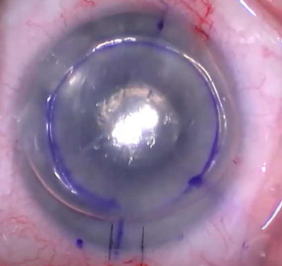
A DSAEK corneal transplant is usually performed under local anesthesia, plus intravenous or oral sedation as needed. Descemet's membrane and the endothelium are replaced with a corneal graft (including a small amount of stroma), but the rest of the cornea remains in place. The graft is obtained from a donor who died recently and is carefully examined to ensure that it is suitable for transplantation.
After anesthetizing the eye, a small incision is made near the edge of the cornea. Through the incision, Descemet's membrane and the endothelium are removed and replaced with the graft. An air or gas bubble is placed under the graft to hold it in place. Sutures are not used to hold the graft in place, but the small incision is sutured closed. If the graft does not adhere sufficiently, a procedure of injecting a repeat air bubble may be needed during the first weeks after the initial procedure.
The "automated" in the name DSAEK refers to the manner of preparing the donor tissue. It is shaved from the donor eye with a microkeratome, a precise oscillating blade.
The advantages of DSAEK compared with a full-thickness corneal transplant
The recovery of the corneal transplant is faster after DSAEK than after a full-thickness transplant, provided that the eye does not have other problems that would limit the potential visual acuity. Although it is quite blurred immediately after DSAEK surgery, the vision usually begins to improve within a month to two months after the surgery. Usually patients can be fitted with glasses within 4 to 6 months (compared with 12 to 18 months after a full-thickness transplant). In addition, because only the thinnest inner layers of the patient's cornea are replaced, the integrity of the cornea is affected less than in a full-thickness transplant.
Partial corneal transplant DMEK
A partial corneal transplant of the Descemet type (DMEK) is the least invasive of the partial-thickness corneal transplants, because the smallest amount of tissue – Descemet's membrane and the endothelium and without stroma – is removed and replaced with a graft of donor tissue. As with DSAEK, DMEK can be used when Descemet's membrane and the endothelium are the corneal layers responsible for vision problems.
How is it performed — DMEK
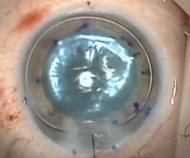
DMEK is usually performed under local anesthesia, plus intravenous or oral sedation as needed. As with the other types of corneal transplant, the corneal tissue graft used in DMEK is obtained from a donor who died after careful tests to ensure that it is suitable for transplantation.
After anesthetizing the eye, a small incision is made near the edge of the cornea. Through the incision, Descemet's membrane and the endothelium are removed and replaced with the graft. An air or gas bubble is placed under the graft to hold it in place. Sutures are not used to hold the graft in place, but the small incision is sutured closed. If the graft does not adhere sufficiently, a procedure of injecting a new air bubble may be needed during the first weeks after the initial procedure.
The advantages of DMEK compared with DSAEK and a full-thickness corneal transplant
The recovery of the corneal transplant is faster after DMEK than after a DSAEK transplant or a full-thickness transplant, provided that the eye does not have other problems that would limit the best potential vision. Although vision is quite blurred immediately after DMEK, the vision usually begins to improve within a week to two weeks after the surgery. Usually patients can be fitted with glasses within a month to two months (compared with 4 to 6 months after DSAEK and 12 to 18 months after a full-thickness transplant). In addition, because only a very thin tissue graft is used for DMEK, the possibility of graft rejection may be lower than in a DSAEK transplant and a full-thickness transplant, and there is a higher chance that the final vision after the surgery can reach 20/20 with glasses.
Risks associated with corneal transplants
As with any type of surgical procedure, a corneal transplant carries some possible risks and complications. Graft rejection is one of the possible risks. If graft rejection or another problem occurs, but the eye retains vision potential, a re-transplant procedure can be performed. The procedure may be identical to the first surgery, an option for a different partial-thickness corneal transplant, or a full-thickness transplant.
Corneal transplant privately
Is it possible to perform a corneal transplant privately?
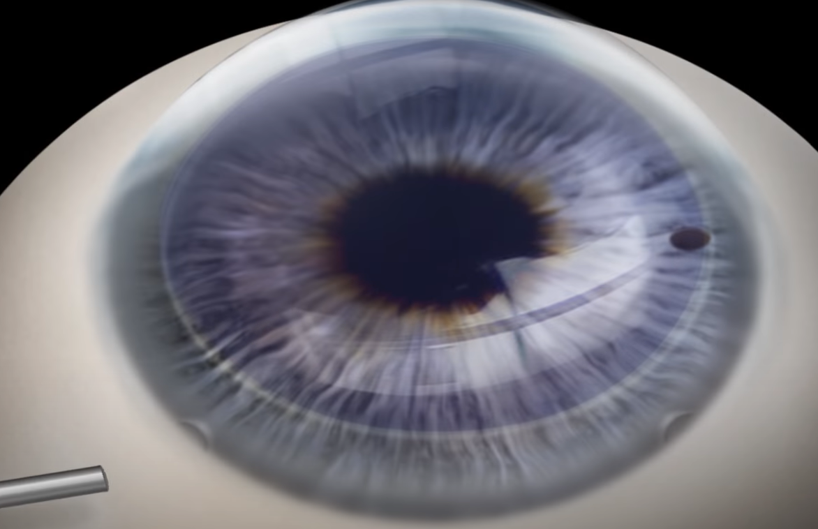
The answer is yes. A corneal transplant can be performed privately and also in a public hospital. Private corneal transplant surgery is made possible because there is an option to import corneas for transplantation from a certified corneal bank abroad. If you have private medical insurance or additional insurance services from your health fund, then they will cover a significant part of the cost of importing the cornea for a private corneal transplant. In addition, if you chose to undergo the surgery with a doctor/center that is in arrangement with them, they will cover the cost of the surgeon's fee and the cost of the operating-room fee.
With me, who should perform a corneal transplant privately?
It is right to perform a corneal transplant privately with a corneal-transplant specialist who is experienced in the surgical method suitable for your eye. Today there are various and advanced methods for performing a corneal transplant and each of them has its own learning curve, and therefore it is advisable to undergo the surgery with someone who has undergone appropriate training for it:
- Full corneal transplant – PKP – in which all the corneal layers are replaced – for example after trauma
- Corneal transplant DALK – in which the innermost/posterior layer is left – for example for keratoconus
- Posterior corneal transplant DSAEK – in which the endothelium, Descemet and a little posterior stroma layer is replaced/transplanted – for example for complex eyes with failure of the endothelium layer
- Posterior corneal transplant DMEK – in which only the endothelium and Descemet layer is replaced – an advanced surgery usually performed in patients with Fuchs dystrophy (FUCHS DYSTROPHY) or those with failure of the endothelium layer after cataract surgery
What does the price of a private corneal transplant surgery consist of?
The price of a private corneal transplant surgery consists of the cost of the cornea, the surgeon's fee and the cost of the operating room at the medical center where you will undergo the surgery. The cost of the cornea and the surgeon's fee vary according to the requirements of the tissue and the surgeon's agreements, and the cost of the operating room varies from one medical center to another according to the type of surgery, the length of the surgery, the required medical staff and the need for the presence of an anesthesiologist. For an exact, up-to-date quote for a private corneal transplant per eye, please contact us.
What is the waiting list for a private corneal transplant?
The waiting list for a corneal transplant in general depends on donor tissue. When undergoing private corneal transplant surgery, because the cornea is ordered from a certified corneal bank abroad, the wait itself is usually a matter of a few weeks depending on the surgeon's availability.
Is there an advantage to performing a private corneal transplant surgery compared with the public system?
As noted, the availability of tissue will be higher when performing a corneal transplant privately, because these are purchased from abroad and matched exactly to the patient's needs. In addition, you will have the option to choose your surgeon and make sure that you get a corneal specialist who is experienced in a corneal transplant with the technique most suitable for your eye.
– Prof. Mimouni performs all types of corneal transplants also in private settings – for details, contact us.
Have a question about your eyes? Want a specialist opinion?