Laser vision correction
There are many methods of laser vision correction, and what they all have in common is the use of an excimer laser and/or a femtosecond laser. All the methods can be divided into two sub-groups:
- Lamellar procedures, such as LASIK
- Surface ablation procedures, such as PRK
Laser vision correction with the excimer laser
The excimer laser is an argon-fluoride laser, and it treats the refractive error (the prescription in your glasses) by performing photoablation of the anterior stroma of the cornea in order to change the radius of curvature of the cornea. In surface ablation methods, the most anterior layer of the corneal stroma — Bowman's layer — is in effect exposed, and the laser is then applied. In the lamellar LASIK method, by contrast, an incision is made in the cornea in order to raise a flap, either with a laser (femtosecond assisted) or with a blade (microkeratome assisted), and the laser is applied to deeper layers of the cornea with minimal damage to the corneal epithelium. Each of the methods has advantages and disadvantages, and not every method suits everyone.
Surface ablation procedures
The surgical methods that belong to this group are (from the most common to the least common): PRK (photorefractive keratectomy), LASEK (laser sub-epithelial keratomileusis) and, least common of all, Epi-LASIK.
PRK (photorefractive keratectomy)
This is the original method of ablating the anterior surface of the cornea for laser vision correction. In this method, after the corneal epithelial layer has been removed (with a laser, with alcohol or mechanically), an excimer laser is applied to the anterior stroma of the cornea in order to change its shape and correct the refractive error. This method underwent multiple clinical trials, including approval by the American FDA, before it reached the wider market. With the invention of LASIK, the use of PRK declined because of the advantages LASIK brings (mainly faster recovery, less pain, and a lower chance of the complication known as haze when correcting high prescriptions with PRK). 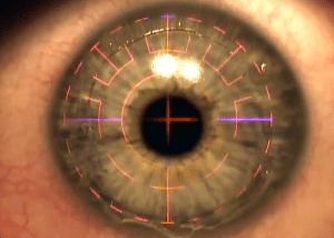
The indications for PRK (as opposed to LASIK) today are: young patients (under 21), thin corneas (under 500 microns), an expected thin residual stromal bed after surgery (under 300 microns), a high percent tissue altered (over 40%), and irregular topography or tomography. All of these are in fact risk factors for ectasia, a very rare complication that can lead to a corneal transplant.
Further indications for PRK are eyes that suffer from diseases of the anterior surface, such as epithelial basement membrane dystrophy or sub-epithelial infiltrates, where the gain here is twofold, since both the refractive error can be corrected and the underlying disease treated. In addition, patients at high risk of a blow to the eye (as part of their occupation or a hobby — a boxer, for example) are best advised to avoid LASIK or any operation involving a flap, since the blow itself can displace the flap. In addition, patients suffering from mild to moderate dry eye are better having PRK rather than LASIK, while patients with moderate to severe dry eye are generally best avoiding laser vision correction altogether without first bringing the dryness under control, since it may worsen after surgery.
You can read more about PRK.
LASEK (laser sub-epithelial keratomileusis)
LASEK is a procedure that is less used today than PRK. It is a method similar to PRK, except that instead of removing the epithelium entirely, it is moved aside in one piece and returned to place at the end. Supporters of this method will say that it speeds up healing of the anterior surface of the cornea and reduces pain, but several large studies that examined this question found no clear advantage for this method over PRK, and sometimes even disadvantages.
Laser vision correction by Epi-LASIK
Epi-LASIK is another method that is less common today. In this method a flap of epithelium alone is created with a special microkeratome that is able to cut a very thin layer, so that the flap consists of epithelium only. Because of the high cost of the microkeratome and the fact that this method has not been shown to reduce pain or speed healing (compared with PRK), this method did not take hold in the market and few perform it.
Lamellar procedures for laser vision correction
The surgical methods that belong to this group of laser vision correction are (from the most common to the least common): LASIK (laser in situ keratomileusis), SMILE (small incision lenticule extraction), ReLEx (refractive lenticule extraction) and FLEx (femtosecond lenticule extraction).
Laser vision correction by LASIK
LASIK (laser in situ keratomileusis) is currently the most common method, because of the fast recovery and minimal pain for patients. This method combines two stages: a first stage of creating a stromal flap, either with an additional laser device (femtosecond) or with a blade (microkeratome); and a second stage of applying an excimer laser to correct the refractive error beneath the flap.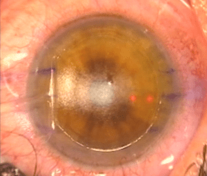 The advantages of this method stem from the fact that there is minimal damage to the corneal epithelium, and so there is no need to wait several days for it to heal. Patients experience minimal pain after surgery, with a fast recovery. Therefore, anyone with no contraindication to LASIK (listed above on this page in the section describing PRK) is most likely better having LASIK than PRK.
You can read more about LASIK.
Laser vision correction by SMILE (small incision lenticule extraction)
SMILE (small incision lenticule extraction) is a relatively new method that came to market in 2009 and received FDA approval in 2016 for myopic patients (−1 to −8) with astigmatism of less than 0.5 dioptres, over the age of 22, and with a refraction that has been stable for over a year.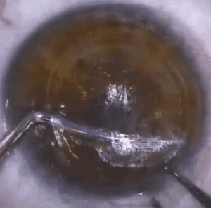
In this method only a femtosecond laser is used, to make two incisions in the depth of the cornea and in effect create a disc that can be extracted through a small incision in the cornea. The advantages of this method are that on the one hand there is no damage to the corneal epithelium, and on the other there is in effect no need for a flap (with all the disadvantages and complications a flap can bring). Supporters of this method will say that it in fact combines the advantages of PRK and of LASIK, but it is still relatively new and is being examined in clinical studies.
You can read more about SMILE.
How do we know who is suitable for laser vision correction?
Patient age and laser vision correction
In the past, because a stable refraction (a stable prescription) was an essential condition for elective refractive surgery, the most widely accepted youngest age was 18–21 years, and the youngest age the FDA approved for laser treatment is 18 years. The accepted minimum age for laser vision correction is 17–18 and above. Although teenagers are indeed the group of patients most eager to have a procedure to correct their vision, the patient's family and the patient themselves must understand that there is an increased chance of needing retreatment, and that a residual stromal bed of at least 300 microns is required, particularly in this group, in order to avoid ectasia. There is no upper age limit for laser vision correction, but the early stages of a cataract in the lens make lens replacement surgery (cataract surgery combined with implantation of an intraocular lens) the preferable option.
Refractive error (the size of the prescription) and laser vision correction
When it comes to small prescriptions, which sometimes go down to as little as 0.5 dioptres (half a unit), LASIK or another procedure can be offered to the patient largely according to their own preference. The problem is with the large prescriptions, where there are disagreements about the prescription up to which laser vision correction can be offered. Most surgeons will agree that the upper limit is up to 10 dioptres for myopia and up to 4 dioptres for hyperopia, depending on the patient. Improvements in technology involving ablation profiles such as wavefront optimized, wavefront guided or topography guided reduce the chance of optical aberrations, glare and haloes after surgery. Even so, excessive use of technology that could lead to an abnormal corneal structure is not recommended, particularly given the availability of further surgical options such as phakic IOLs. Astigmatism of up to 5 dioptres can be treated, while for larger amounts of astigmatism, with sufficient corneal thickness, a staged treatment can be used. If there is a discrepancy between the subjective refraction, the topography and the keratometry, lenticular astigmatism has to be taken into account and the treatment adjusted according to a weighted average, based on various clinical considerations for each case individually.
Corneal thickness and laser vision correction
Normal corneal topography is an essential condition before excimer laser vision correction is performed. LASIK must under no circumstances be performed in eyes with features of early keratoconus or pellucid marginal degeneration, or that are suspected of these diseases.
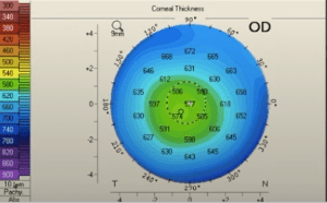
The reason for this is that eyes with these features are at increased risk of ectasia after surgery. There are grading systems that make it possible to assess the risk of ectasia after LASIK. If there are mild irregular findings on topography (for example mild inferior steepening), a surface ablation procedure can be offered. In any event, if there is doubt, it is better to avoid corneal surgery and better to perform lens-based surgery.
Keratometry and laser vision correction
Keratometry matters because the cornea is flattened in order to correct myopia and steepened in order to correct hyperopia. In general, a minimum value of 34 dioptres and a maximum value of 50 dioptres after surgery are desirable, and this must be calculated before the operation. Beyond these values there may be degradation of optical quality.
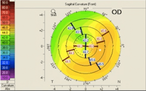
There are many topographic methods that use the keratometry of the map in order to screen for keratoconus. One classic example of such a method is the I-S ratio, or inferior-superior ratio, of Rabinowitz. This method describes a ratio of 1.9 and above as consistent with keratoconus, and a range of 1.4–1.9 as suspicious for keratoconus.
Pupil size and laser vision correction
No correlation has been found between pupil size and subjective complaints of aberrations, glare or haloes. Even so, patients with very wide pupils under mesopic conditions (7.5–8.0 mm) are most likely at increased risk of optical aberrations. The FDA listed a wide pupil size in dim light as a possible warning sign against performing LASIK, and so such a finding must be discussed if it is identified in a patient. The new algorithms of the laser devices produce fewer optical aberrations, because there are better transition zones (the transition between the treated zone and the untreated zone) with aspheric curves, and so pupil size is becoming less critical.
What is important to ask every patient?
A detailed history of the patient before surgery is a critical part of the process, including a complete ocular history. Patients with a background of ocular herpes, strabismus, double vision, previous refractive surgery, dry eye or contact lens intolerance have a lower chance of successful refractive surgery.
A history of ocular herpes simplex virus — is laser vision correction permitted?
Animal experiments have demonstrated that the excimer laser can cause a recurrence of ocular herpes. Clinically, there have been reports that LASIK, PRK and even PTK led to a recurrence of ocular herpes. Many surgeons therefore advise patients with a background of ocular herpes, even if it is not active, to avoid LASIK or PRK. Even so, some patients with a background of ocular herpes will still prefer to have the surgery for personal reasons (an occupational need, for example). In such patients the surgery can be considered, but it is important to make clear to them the increased risk of complications after surgery, and it is highly advisable to consider prophylactic oral antiviral treatment before and after the operation.
Does a history of childhood strabismus rule out laser vision correction?
Patients with a history of childhood strabismus have in the past developed a recurrence of the strabismus after LASIK. Patients who wear prisms in their glasses should be aware that they will most likely need prisms after laser vision correction as well. Patients with a history of strabismus with a preference for fixation in their dominant eye may not get on with monovision, since they may find it difficult to switch fixation to their non-dominant eye for reading.
Is laser vision correction permitted in monocular patients — a single seeing eye?
It is advisable to recommend that patients with poor vision in one eye avoid elective laser vision correction. Although the chance of a serious complication is low, it still exists. Some surgeons will say that if the patient says they can function with the weaker eye alone (mild amblyopia), the surgery can still be considered. On the other hand, there may be cases in which patients with very poor vision in one eye still insist on having the surgery. In these cases, if there is agreement on both sides to proceed, it is very important that there is a thorough discussion of the risks involved.
Can people prone to keloid formation have the surgery?
Although a history of keloid formation in the skin was once thought to be a risk factor for corneal haze after surface ablation, numerous reports have demonstrated that LASIK and PRK do not put patients with a history of keloids at risk.
Can people who wear contact lenses have laser vision correction?
Patients who wear hard lenses should be warned that it sometimes takes weeks to months until the cornea returns to its normal shape, and so they may need repeat examinations (until they are shown to be stable) in order to determine whether they are in fact suitable for laser vision correction. There are studies that have demonstrated very clearly that patients with dry eye before surgery are at increased risk of the dryness worsening after LASIK.
Are patients with presbyopia (needing reading glasses) suitable for laser vision correction?
Presbyopic patients (who need reading glasses) should be questioned and assessed to see whether they are suitable for monovision. Since some patients are unable to adapt to monovision, it is important to confirm with the patient that they have got on with monovision in the past, or to give them the opportunity to try contact lenses themselves as a monovision feasibility test, to check that they are indeed able to adapt.
Can pregnant women have laser vision correction?
Patients who are pregnant, who have recently given birth or who are still breastfeeding should be warned that in their case laser vision correction may lead to sub-optimal results.
Which laser vision correction procedure is recommended for people who take part in higher-risk physical activity?
Patients who take part in physical activities with an increased risk of trauma to the eye area, such as martial arts (karate, boxing, judo and so on), extreme sports or any other activity with an increased risk of trauma to the eye area, are at higher risk of flap dislocation after LASIK. A laser procedure using surface ablation, such as PRK, can therefore be considered in such patients.
Which medications can interfere with the results of surgery?
There is a wide range of medications that may affect the results after refractive surgery. For example, Roaccutane (a treatment for acne) can lead to severe dry eye over a long period. Laser vision correction should therefore be avoided in such patients for as long as they are being treated with this drug, and it is worth waiting for a period after the drug is stopped in order to allow the dryness to resolve.
Which medical conditions can interfere with healing after surgery?
There are systemic medical conditions that can affect healing after surgery. Diabetes, immunosuppressed states such as HIV, and autoimmune conditions such as lupus can all impair the results of surgery, because these conditions alter the body's wound-healing process. Even so, several studies have recently reported success with LASIK in patients with autoimmune diseases, and so, if they are completely stable with regard to their underlying disease with no ocular manifestation, and it has been explained to them that there is an increased risk, surgery can still be considered.
The risks of laser vision correction
What are the possible risks and complications of laser vision correction?
- The safety profile of flap-related complications after LASIK has improved significantly over the past decade. Even so, laser vision correction does carry risks.
- Intraoperative LASIK complications can often be prevented with the appropriate safeguards of the surgical system.
- Femtosecond flaps provide a planar cut of more reliable and uniform thickness and greatly reduce the incidence of flap striae.
- The incidence of diffuse lamellar keratitis (DLK) and diffuse infections falls with the use of the femtosecond laser.
- The use of potent topical steroids has reduced the need to lift and irrigate flaps as a treatment for complications such as DLK.
Introduction to the risks of laser vision correction
Understanding the difference in perspective between a patient undergoing LASIK and a patient undergoing cataract surgery is critical to understanding what is distinctive about these patients and their expectations. Cataract patients must have cataract surgery or they will continue to lose vision, so they are prepared to accept some risk in their procedure. Patients undergoing LASIK have no disease and no loss of vision, and so any complication comes as a surprise and causes enormous anxiety, regardless of how well informed they were before surgery. Given this perspective of a patient undergoing LASIK, the risk of complications must be particularly low (<1/1000 for significant complications requiring further procedures). Fortunately, improvements in LASIK techniques and technology over the past two decades have allowed us to achieve this high safety profile.
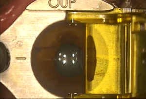
A worldwide literature review of LASIK completed in 2008 showed an overall satisfaction rate of 95.4%, with the most common reasons for dissatisfaction being residual refractive error, dry eye, older age and symptoms related to disturbances of night vision. LASIK complications can readily be divided into complications that occur during surgery (intraoperative), in the early postoperative phase and in the late postoperative phases. Most LASIK complications can be corrected so that there are no long-term consequences. Even so, there are a few rare complications that can have permanent consequences for vision.
Intraoperative LASIK complications (complications that can occur during surgery)
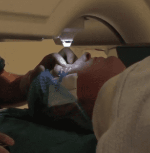
Intraoperative LASIK complications can often be prevented with the appropriate safeguards of the surgical system. If the equipment is faulty or incorrectly configured, a complication is all but guaranteed. The key to minimising LASIK complications is therefore prevention. Proper training and a high degree of focus are essential for both the surgeon and the surgical technicians in order to minimise surgical errors.
The LASIK flap, although traditionally created with a microkeratome (the blade method), is now increasingly performed with the femtosecond laser (a femtosecond flap). There are advantages and disadvantages to both approaches.
Risks and complications in creating a flap with a femtosecond laser
The femtosecond device works using photodisruption technology, allowing a laser focused for a short time to split tissue by means of thousands of bubbles (cavitation). These cavitation bubbles, when they overlap slightly, can create precise planes of tissue in many different patterns. Femtosecond lasers therefore create planar flaps of uniform thickness and allow more predictable biomechanical changes in the tissue.
Suction loss during flap creation
The rate of suction loss is very low, at around 0.006%, although it is generally more common among less experienced surgeons. It usually happens at the start of the learning curve of docking the equipment before performing the cut itself. There are a variety of causes, such as deep-set eyes, a prominent forehead, a narrow palpebral fissure, a very flat cornea, forceful eyelid closure by the patient, and the patient's inability to maintain fixation or follow instructions. The most common causes of suction loss are excessive movement of the suction ring on the laser cone and movement of the patient's head while the patient is docked under the femtosecond laser.
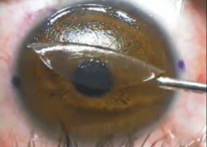
The loss of the meniscus will be visible on the screen, and the loss of suction may also be felt by the surgeon. A smaller meniscus will appear on the screen. Movement of the patient's head can be observed during the interruption of suction. Vertical gas breakthrough can develop if application of the laser continues despite the loss of suction.
Managing suction loss during flap creation
The stage of the lamellar cut at which the suction loss occurs dictates how it is managed. When suction loss occurs at the initial stages, before the sidecut has been made, the flap can be repeated at the same diameter with minimal change (cancelling the pocket).
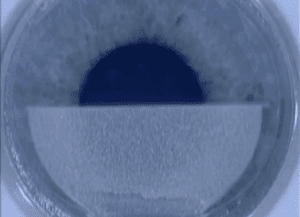
It is worth using the same cone in order to ensure that the second cut is made at the same depth, but it is worth changing the suction ring in order to improve the suction on the second attempt. It is best not to wait too long (more than 10 minutes) between attempts, or conjunctival oedema may develop that will interfere with a further attempt. When suction loss occurs just before the start of the sidecut, or while it is being made, the raster stage (the lamellar cut of the flap) can be skipped and the sidecut made at a diameter 0.5 mm smaller than on the previous attempt.
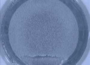
It is important that the flap is lifted from the furthest point at which the surgeon is confident the correct plane has been found. It is best not to lift the flap in the area where the suction loss occurred, since an incomplete flap lift may result. A further alternative is to wait 1–3 months and perform advanced surface ablation (PRK). PRK can also be performed immediately after an unsuccessful flap has been created, as long as the flap has not been lifted.
Preventing suction loss during flap creation
Suction loss can be minimised by ensuring adequate suction before starting to apply the laser. In general, a suction reading of less than four on the syringe of the IntraLase laser indicates adequate suction. During the docking process, the surgeon should try to keep the plane of the suction ring perpendicular to the line of descent of the laser cone in order to minimise movement of the suction ring. Patients should be encouraged to stay still in order to minimise movement of their head.
Vertical gas breakthrough
Femtosecond lasers create a lamellar plane with plasma separation. If there is a break or a scar in the anterior stroma, gas can escape from the lamellar plane through that scar and accumulate on the anterior surface of the cornea. This anterior gas bubble will then block the subsequent laser pulses, resulting in an incomplete flap in the area of the gas bubble.
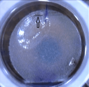
The gas breakthrough will appear as a clear or black round spot on the grey lamellar plane of the flap.
Managing vertical gas breakthrough
Small bubbles of gas breakthrough (<1 mm) will usually still allow the flap to be separated and lifted when rapid dissection is carried out through the interface in the area of the breakthrough. Larger areas of gas breakthrough (>1 mm) will not allow the flap to be separated. As long as the flap has not been lifted, the surgeon and the patient have the option of switching to a PRK procedure. Since additional trauma has been caused to the cornea by the femtosecond laser, topical mitomycin should be considered.
Preventing vertical gas breakthrough
Patients should be examined for stromal scars before femtosecond LASIK is recommended. Scars that are recent (<2 years), larger (>1 mm) and deeper (>20% of corneal thickness) will generally be associated with vertical gas breakthrough and the creation of an incomplete flap. Patients with scars should be informed of the risk of gas breakthrough and given the option of having primary PRK, or of attempting femtosecond LASIK on the understanding that their procedure may need to be changed to PRK later. Increasing the flap depth by 20 microns may also reduce the risk of gas breakthrough, by creating the flap interface beneath the scar.
Gas bubbles in the anterior chamber
With the femtosecond laser, gas can rarely escape into the anterior chamber. The mechanism is most likely that air can occasionally pass through Schlemm's canal into the anterior chamber, although this is not supported by optical coherence tomography (OCT) imaging. Bubbles in the anterior chamber occur more frequently when the edge of the flap is close to the limbus, or if there are blood vessels in the peripheral cornea. It has previously been reported that anterior air bubbles arising from the femtosecond laser do not affect endothelial cell density compared with normal eyes. The bubbles can affect tracking on certain lasers, by making it difficult for the laser to locate the centre of the pupil and the centre of the treatment.
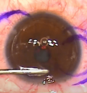
Managing gas bubbles in the anterior chamber
The surgeon can wait until the bubbles are reabsorbed (although this may take more than 4 hours), or continue with the excimer correction using manual centration after switching off or disabling eye tracking. While other surgeons suggest dilating the pupil in order to achieve tracking, this will still lead to a disturbance in centration because of the decentred pupil that follows dilation, and so this technique is not recommended.
Preventing gas bubbles in the anterior chamber
For patients with small corneas, the flap size can be reduced by 0.5 mm in order to reduce the risk of bubbles in the anterior chamber. In patients with significant blood vessels around the periphery of the cornea, PRK rather than femtosecond LASIK can be considered.
Opaque bubble layer
If gas bubbles are not allowed to pass uniformly through the lamellar interface, into the pocket and then out of the treatment area, they will accumulate in the stroma of the cornea and cause an opaque bubble layer (OBL). The OBL will cause adhesions at the lamellar interface that make the flap harder to lift, and can also prevent the infrared-based eye tracker from obtaining a clear image of the pupil. OBL of the peripheral cornea can reduce the accuracy of iris registration by obscuring the limbus. Mild adhesions can be separated carefully, but attempts to separate larger or more cohesive adhesions may cause a tear in the flap. The effect of OBL on the excimer treatment itself is unclear; however, it does not appear to change the results of treatment.
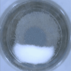
Managing the OBL complication
OBL can be reduced by gently rubbing the stromal bed with a smooth instrument such as a cannula. Surgical spears should not be used, since they will dry the cornea and alter the effects of the excimer laser. The infrared tracking monitor allows the surgeon to follow and identify when enough OBL has been removed for accurate tracking to be achieved. Alternatively, the surgeon can wait 10–30 minutes until the OBL is absorbed into the cornea and then proceed with laser vision correction without risk.
Preventing the development of OBL in laser vision correction
The key to avoiding OBL is creating a good meniscus during the docking process. A good meniscus (halfway between the edge of the cone and the edge of the flap) comes from good docking technique. With a good meniscus, there is minimal risk of OBL forming. Avoiding excessive pressure, which results in a smaller meniscus, can also prevent OBL from forming. An excellent meniscus at the time of docking should touch the edge of the pocket; this allows the gas to escape into the pocket. "Soft docking" is another technique used to reduce OBL. In this technique, once applanation has been achieved, the laser cone is raised slowly in order to increase the size of the meniscus down to minimal applanation.
Complications and risks in creating a flap with a microkeratome
Before a LASIK procedure, all the equipment should be checked, since any equipment fault could cause a flap complication. The rate of all flap complications has been reported at around 0.3%, with failure to achieve intraocular pressure in 0.034%, partial flaps in 0.099%, a buttonhole in 0.07%, thin or irregular flaps in 0.087%, and free caps in 0.012% of eyes. Microkeratome blades supplied by the manufacturer may be defective. Defects or irregularities in the cutting edge will produce an abnormal flap. It is difficult to inspect the microkeratome blade once it has been loaded into the microkeratome, since the edge is not well reflected by the microscope light. If a microkeratome pass is made with an immobile blade, an irregular flap will be created. A test should be run with the microkeratome to confirm that it moves smoothly along the microkeratome track.
Buttonhole formation and an irregular flap with a microkeratome
A buttonhole occurs when a microkeratome pass has been made without adequate suction. Steeper corneas (above 49 dioptres) most likely also carry an increased risk, although there are conflicting reports in the literature. The formation of a buttonhole is apparent immediately after the microkeratome has completed the pass. The defect through the centre of the flap will be obvious. The stromal bed has a clear area at the centre, about 2–3 mm in size and slightly raised, representing the uncut area of cornea where the epithelium is still present. After a month, haze will form around the edges of the hole.
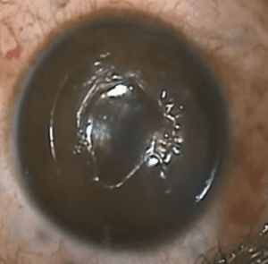
Managing a buttonhole in the flap
Performing laser ablation will cause irregular astigmatism with haze in the central cornea. If the hole is identified immediately, the flap should not be lifted. Gentle irrigation of the interface will allow the flap to be placed back in position. If the flap has been lifted, it should be returned to its place. Correct positioning of the flap may be difficult to determine, since the flap is thin and very unstable. The eye should be allowed to heal for at least several weeks to months before attempting any further refractive correction. Topical steroids should be used during the healing phase in order to reduce the risk of haze forming. Immediate trans-epithelial PRK can be offered following flap complications, but this should only be done after careful discussion with the patient.
Preventing buttonhole formation
A buttonhole in the flap can be prevented with the techniques and precautions discussed. Most important of all is to avoid making a microkeratome pass without adequate suction.
Have a question about your eyes? Want a specialist opinion?