Keratoconus
Keratoconus of the cornea is a disease that occurs in 0.05% of the population. In this disease the central or paracentral part of the cornea undergoes thinning and bulging, so that the cornea takes on the shape of a cone (which is where the name comes from).
The familial, genetic or environmental component of keratoconus

On the one hand, this disease has a genetic component: between 5 and 10% of patients report a family history. On the other hand, it is not possible to predict who will develop the disease, and in 90% of patients there is no family history. It is worth noting that studies have shown that relatives of keratoconus patients, even if they are asymptomatic, tend to have sub-clinical thinning of the cornea. Researchers have identified a number of chromosomal regions associated with a higher risk of developing the disease, but specific genes have not yet been identified. The prevailing view today is that the disease arises from a combination of several factors (multifactorial), such as family history, excessive eye rubbing, chronic inflammation or chronic irritation of the ocular surface. There are several conditions in which there is a higher risk of developing keratoconus; in a large proportion of them the mechanism is most likely a tendency to rub the eyes. These include Down syndrome, Marfan syndrome, floppy eyelid syndrome and various congenital eye conditions.
The typical histological appearance of keratoconus
When the histology of corneas affected by the disease in its early stages is examined, fibrils can be seen appearing in Bowman's layer, leading to damage and "breaks" in that layer. When the stroma and the epithelium above it are examined, mainly thinning and/or diffuse scarring can be identified. In Descemet's layer, folds and breaks can be identified. An inflammatory process is not usually identified in the various layers.
Clinical signs of keratoconus
In most cases the disease appears, or is partly expressed, in both eyes. There may be a situation in which the disease appears in a far more severe degree in one eye. The disease usually progresses and worsens during adolescence and continues into the thirties, but it can appear at any age. There are several clinical signs that can be identified in this disease.
Scissoring of the red reflex
This is an examination performed with an ophthalmoscope, in which the normal red reflex shows something like two blades of a pair of scissors moving apart and together.
Rizzutti sign
A pen torch is used to shine light on the cornea from the temporal side, and a cone-shaped reflection of light is produced on the nasal side.
Munson sign
This appears at a slightly later stage. The patient is asked to look down and is observed from above, when a bulge of the lower eyelid can be seen.
Assessment and follow-up of patients with keratoconus
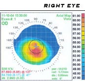
Corneal topography, or computerised videokeratography, makes it possible to identify the disease at early stages, to follow its progression by comparing examinations over time, and to fit the patient with contact lenses according to their topography. When these devices are used it can be seen that in most patients the bulge itself appears mainly on the lower side of the cornea (inferior steepening), while the thinning of the cornea is in a ring around the centre (paracentral thinning).
Treatments for keratoconus
In the early stages of the disease it is sometimes enough to use glasses to correct the astigmatism created by the cone. Even so, the preferred treatment as early as the fairly early stages is rigid contact lenses, which in a large proportion of cases succeed in neutralising the cone and the astigmatism it creates, leading to a marked improvement in patients' quality of vision.
Penetrating keratoplasty in keratoconus
Indications for surgery
There are several indications for surgery in patients with keratoconus. The first indication is a quality of vision that is inadequate despite a properly fitted contact lens, which usually results from scarring in the various layers of the cornea. The second indication is that the patient is unable to tolerate the sensation of the fitted rigid contact lenses. Progression of the disease, expressed as continued thinning of the periphery approaching the limbus, is a further indication, because the closer the thinning comes to the limbus, the larger the corneal graft the patient will need, with an increased chance of surgical complications. Acute hydrops is not an indication for immediate surgery.
Surgical options and prognosis
The classic operation is PKP (penetrating keratoplasty), in which a corneal graft containing all the layers of the cornea is transplanted. Successes have recently been reported with DALK (deep anterior lamellar keratoplasty), in which all the layers except the endothelium are replaced in order to reduce the chance of graft rejection. In any event, the prognosis after surgery in keratoconus patients is excellent.
Cross-linking — a treatment for keratoconus
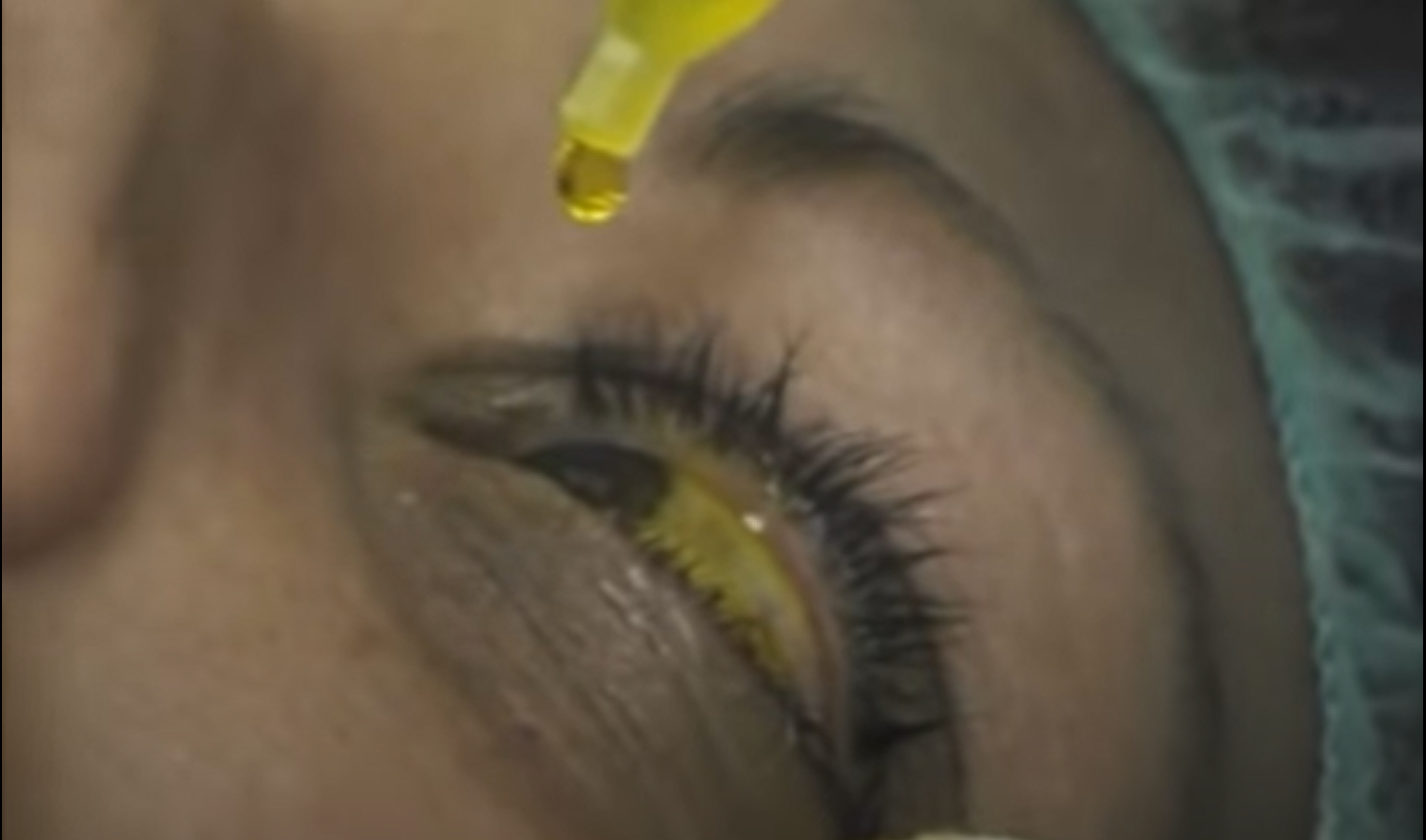
This is a new method of preventing progression of the disease by strengthening the mechanical strength of the layers of the cornea. In this method riboflavin is instilled into the eye and UV-A light is then applied for 30 minutes. The riboflavin causes new bonds to form between the collagen fibres within the stroma of the cornea.

Intrastromal corneal ring segments
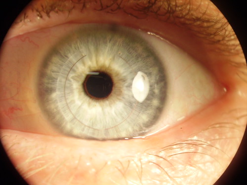
These are rings 210 to 450 micrometres thick that are implanted between the different layers of the stroma. These rings flatten the cornea, and the mechanical change corrects the patient's astigmatism.
Refractive procedures
Keratoconus is a contraindication to refractive procedures such as LASIK or LASEK (commonly known as laser vision correction). The reason is that keratoconus patients who undergo such a procedure will have poor results in the short term and even more so in the long term.
It is important to note that it is well worth consulting an ophthalmologist about all the treatment options for keratoconus and deciding together which treatment is best for you.
Is there an effective natural treatment for keratoconus?
There is as yet no natural treatment for keratoconus that can be taken into the eye or by mouth that will halt the progression of keratoconus or improve vision in patients with damage from keratoconus.
What can be done to halt the progression of keratoconus?
Risk factors for keratoconus can be identified, and an attempt made to intervene in those areas of life and those habits. For example, avoiding eye rubbing, and sleeping on your back or with an eye shield in patients with particularly lax eyelids (floppy eyelid syndrome).
Is there a link between eye rubbing and keratoconus?
There is a prevailing view among cornea specialists that patients who rub their eyes, particularly those who do so with their knuckles, are at increased risk of progression of keratoconus. For those patients it is important to identify the reason for the eye rubbing. If it is a habit they cannot control (similar to patients with Tourette syndrome or severe OCD), then a change in habits should be encouraged, along with work on the conditioning that leads to the eye rubbing. If it is eye rubbing that arises from itching in the eyes (seasonal allergy, for example), then anti-allergy eye drops (antihistamines, for example) can be given, which will reduce the sensation of itching and help the patient avoid rubbing.
Is there a link between sleep habits and keratoconus?
It has been identified that there is a particular group of keratoconus patients in whom there is a link between sleep habits and the development of the disease. These are usually people who are overweight with particularly lax eyelids and a tendency to sleep apnoea during the night. Sometimes their partners will report that they snore heavily at night, and they tend to be the kind of people who like to sleep hugging a pillow right up against their face.
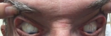
For those patients we recommend first of all that their family doctor refers them to a sleep laboratory, because it is now known that there is a link between sleep apnoea and lax eyelids (floppy eyelid syndrome). Sleep apnoea has consequences for the whole body beyond the eyes, and so it is important to refer them for this examination. In addition, patients can be advised to sleep on their back, or with a clear plastic shield or protective goggles when they go to sleep at night, in order to avoid uncontrolled rubbing during sleep.
I was offered cross-linking, but "I want a natural treatment for keratoconus, not chemicals"
Cross-linking has genuinely revolutionised the treatment of keratoconus. 90% of the time this treatment can prevent the progression of keratoconus, and far fewer people need a corneal transplant as a result of this disease. As for the treatment itself: after the outer layer of the cornea (the epithelial layer) has been removed, vitamin B2 — also called riboflavin — is instilled onto the cornea for about 20 minutes. This is admittedly a surgical procedure, but the substance itself (vitamin B2) certainly occurs in nature, in foods such as milk, cheese, eggs, vegetables, mushrooms and others. Once the instillation of the substance is complete, ultraviolet A light is applied to the cornea for a few minutes, which activates the riboflavin and causes cross-links to form between the collagen fibres in the cornea, thereby strengthening it.
Prof. Michael Mimouni is a specialist in the treatment of keratoconus.
Have a question about your eyes? Want a specialist opinion?