What is DMEK?
A DMEK-type corneal transplant, short for Descemet Membrane Endothelial Keratoplasty, is a partial-thickness corneal transplant procedure that includes selective removal of the Descemet membrane and endothelium, followed by transplantation of the donor's corneal endothelium and Descemet membrane without additional stromal tissue from the donor.
How did DMEK develop?
In the last two decades there has been significant progress in corneal-transplant techniques. The treatment of corneal edema resulting from dysfunction of the endothelium developed from a full-thickness corneal transplant
to replacement of the damaged layer alone. This change from penetrating keratoplasty to posterior lamellar surgery lowered the rejection rate after the keratoplasty and preserves the structural integrity of the eye. There are two common posterior partial corneal-transplant techniques today, and they are DSAEK and DMEK.
In 2006 Michael Tappin reported the use of dedicated cannulas to transfer discs of the endothelial layer and Descemet membrane with a diameter of 7.5 mm, which were attached to the exposed stroma of the recipient's cornea by means of air injected into the anterior chamber. Corneal edema improved in the three cases reported by means of this procedure, which was called True Endothelial Cell Transplantation. In parallel, Gerrit Melles presented a similar idea which he called DMEK, a procedure that was later widely adopted by corneal surgeons around the world.
When is DMEK performed (indications)?
The indications for DMEK are similar to those of DSAEK and include endothelial dystrophy (such as Fuchs corneal dystrophy and posterior polymorphous corneal dystrophy), pseudophakic bullous keratopathy (PBK), ICE syndrome and other causes of dysfunction of the corneal endothelium.
What are the advantages of DMEK?
The most significant advantage of DMEK is the fast visual rehabilitation. Moreover, it has the fastest visual rehabilitation compared with any keratoplasty technique to date. The final visual acuity can be exceptional because of minimal optical-interface effects, since less tissue is transplanted (less than 20 micron). In addition, as a result of the minimal amount of tissue, there is a lower risk of graft rejection and less long-term dependence on local steroids compared with other types of keratoplasty. Stopping local steroids can be considered a year after the procedure, or switching to a weaker steroid, especially for patients with elevated intraocular pressure.
What are the disadvantages of DMEK?
Because of its thinness, fragility and the rolling properties characteristic of it (when the endothelium faces outward), the donor tissue can be difficult to handle and contribute to technical difficulties during the surgery. This surgery has a learning curve and experience and skill are required in order to perform it. In addition, even in skilled hands, there is a higher risk of graft detachment compared with DSAEK, and sometimes a bubble-injection procedure is required in the first weeks after the surgery (in 5 to 25% of cases, and this is actually considered part of the surgery).
What are the stages of DMEK corneal-transplant surgery?
1. 2 to 4 small incisions are made in the cornea (less than 1 mm) and a main incision of 2.4 to 3.0 mm in size
2. The anterior chamber is filled with viscoelastic gel
3. A peripheral inferior iridotomy is created using a 30 needle or a reverse Sinskey hook in order to prevent pupillary block (some skip this stage and some perform it a few weeks before the surgery)
4. The recipient's corneal epithelium is marked with a round ring slightly larger than the diameter of the planned graft to create a template for peeling the recipient's Descemet membrane
5. Scoring of the recipient's Descemet membrane in a circular manner using a reverse Sinskey, and then peeling the Descemet membrane from the stroma above it
6. Removal of the viscoelastic material by means of irrigation and aspiration
7. Injection of miochol to constrict the pupil and BSS to normalize the pressure
8. Inserting the tip of the glass tube into the main corneal incision and injecting the donor tissue into the anterior chamber
9. Releasing fluid from the small corneal incisions to shallow the anterior chamber
10. Tapping gently on the surface of the cornea until the graft is properly positioned and unfolding it
11. Injecting air or SF6 gas into the anterior chamber in order to attach it to the recipient's stroma
12. Suturing the main incision with a 10-0 nylon suture (not mandatory if it does not leak at the end of the surgery)
13. Making sure the patient lies supine with the face upward for two hours and checking the patient about two hours after the surgery to make sure there is not too little or too much air/gas in the anterior chamber
-
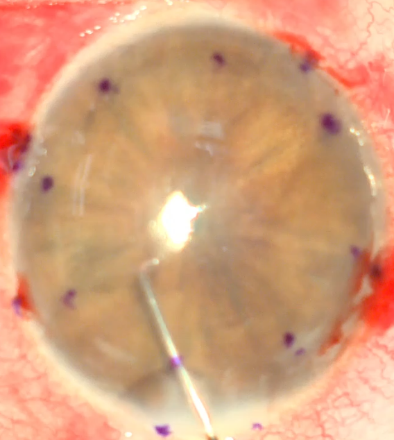
Peeling the Descemet membrane from the recipient -
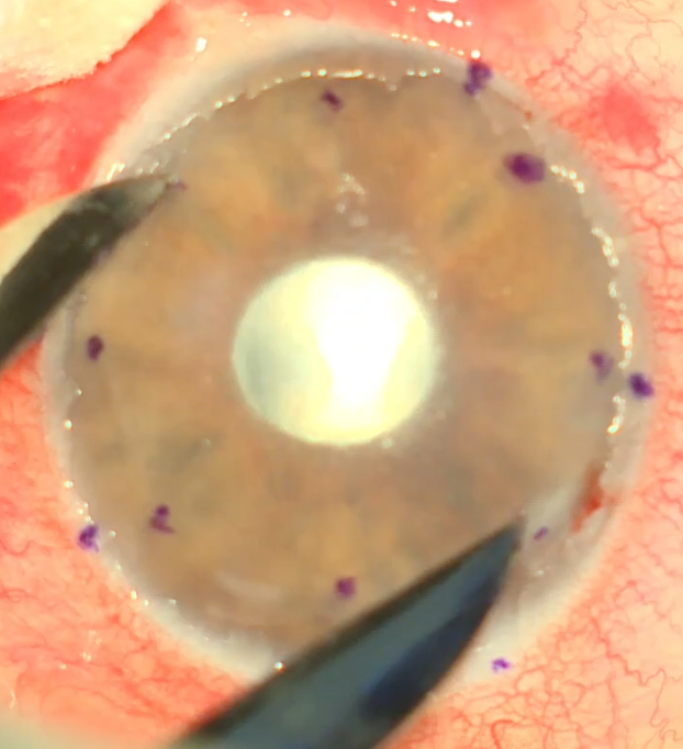
Marking the peeling boundaries -
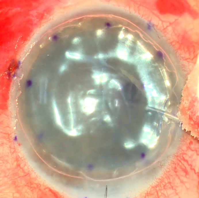
Injecting air into the anterior chamber -
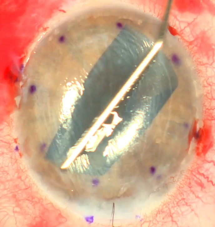
Tapping on the cornea in order to open the graft and unfold it
What is the treatment after DMEK surgery?
The surgery requires frequent visits to the doctor. It is customary to check two hours after the surgery, a day, a week, two weeks, a month, three months and a year after the surgery. After the surgery, antibiotic and steroid drops are given. The antibiotic is stopped a week after the surgery (or until the patient's epithelium heals). In the first days after the surgery there is a need to lie supine on the back during most hours of the day (the first 48-72 hours). Steroid drops are continued at a full dose in the first month after the surgery and are then gradually reduced each month. It is recommended to continue a low dose of steroid drops until at least a year after the surgery. Afterwards, one can consider stopping the steroids or switching to a weaker steroid. During these visits it is important to monitor the patient's intraocular pressure and to make sure there is no response to the steroids leading to a rise in intraocular pressure. As noted, in the first weeks after the surgery about 5 to 25% of patients will need a repeat injection of air into the anterior chamber, and sometimes more than once.
What are the complications of DMEK surgery?
The most common complication of DMEK corneal-transplant surgery is graft detachment that requires a repeat injection of an air bubble (5-25%). In fact, it is not really a complication but part of the surgery, since it is quite a common phenomenon.
Primary graft failure. For a reason that is not always known, in 1-5% of patients there will be no clearing (a decrease in edema) of the cornea after the surgery. Such a state is called primary graft failure. In such a state one can repeat the DMEK surgery or switch to another surgical method such as DSAEK or PKP.
Graft rejection. Graft rejection can occur at any given time after the corneal transplant. Usually starting from a few months up to a few years after the surgery. The chances of experiencing graft rejection can be reduced by a gradual decrease in steroid drops after the surgery and prolonged use of them at a low dose. If there is graft rejection, the dose and strength of the steroids are increased temporarily until the rejection is brought under control. Sometimes there is a need to combine additional treatments in order to overcome the rejection. If graft rejection is treated later, it may lead to secondary graft failure and a need for repeat surgery.
Infections. In less than 0.1% of cases there may be an intraocular bacterial/fungal infection or an infection in the cornea after DMEK surgery. In order to prevent such a state, the surgery is performed in a completely sterile condition and antibiotic drops are used in the first week after the surgery or until closure of the corneal epithelium. In addition, there is a risk of activation of herpes disease after a DMEK transplant, which can present like graft rejection. Therefore, if there is a clinical picture of graft rejection that does not respond to steroids, one must take into account activation of herpes or another intraocular infectious process.
A rise in intraocular pressure. As noted, usually in response to prolonged use of steroids. The treatment is usually a combination of reducing the dose of steroids and adding treatment to lower the intraocular pressure. Another anti-inflammatory substitute that does not raise intraocular pressure can nevertheless be given in order to prevent graft rejection in such situations.
When does one see well after DMEK surgery?
In the first days after the surgery the vision is very blurred because there is an air bubble in the anterior chamber that attaches the graft in place and covers the pupil, so the vision is very blurred. In addition, usually a few days/weeks are required until the corneal edema is absorbed and the cornea begins to be transparent. In parallel, if there was a need to remove the corneal epithelium during the surgery, between a week and two weeks are required for healing of the corneal epithelium. If there is detachment of the graft (as happens in 5-25% of cases), then there is a need for an additional injection of air into the anterior chamber, which slows visual recovery. Usually, about a month to three months after the graft is completely attached, an update of the glasses can be performed in order to understand what visual acuity has been achieved. For comparison, after PKP surgery a recovery period of over a year is required in order to understand what visual acuity has been achieved.
Have a question about your eyes? Want a specialist opinion?