What is a chalazion?
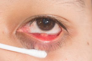 Chalazia (the plural of chalazion) are the most common inflammatory lesions of the eyelid. They usually enlarge slowly and present as painless lumps in the eyelids. A chalazion is usually benign and resolves on its own, although they can cause chronic complications. A chalazion that recurs again and again in the same place should be examined, because it may be a malignancy. A chalazion is a chronic sterile lipogranuloma. A deep chalazion is caused by inflammation of a tarsal meibomian gland. A superficial chalazion is caused by inflammation of a gland of Zeis. In this article we will describe the causes, pathophysiology, presentation and diagnosis of a chalazion.
Etiology
A chalazion is caused by inflammation and obstruction of the oil glands of the eyelids. While an infection may cause the inflammation or the obstruction leading to a chalazion, the lesion itself is an inflammatory and not an infectious lesion.
Epidemiology of a chalazion
This is a common condition, although the exact incidence in Israel or worldwide is not documented. It appears to affect males and females equally, but exact numbers are not available. Chalazia appear at higher frequency in adulthood (ages 30-50).
Pathophysiology
A chalazion is an inflammatory lesion that forms when lipid breakdown products leak into the surrounding tissue and provoke a granulomatous inflammatory response. For this reason, a chalazion is also called a granuloma of the conjunctiva. Meibomian glands are located in the tarsus of the eyelids (the inner part of the eyelid); therefore, edema due to obstruction of these glands usually involves the eyelid-margin area. Occasionally, a chalazion may grow and break through the tarsus to the outer part of the eyelid margin. A chalazion due to obstruction of the glands of Zeis is usually located along the eyelid margin.
Histopathology of a chalazion
Histopathological evaluation is rarely required in the diagnosis and management of a chalazion. Histological examination reveals a chronic granulomatous response with lipid-laden giant cells of the Touton type. Usually, the nuclei of these cells are located around a central foamy cytoplasmic area containing the ingested lipid material. Mononuclear cells, including lymphocytes or macrophages, may also be found at the periphery of the lesion since it is an inflammatory process. If a secondary bacterial infection develops, then one may expect to find an acute necrotic response with PMN-type cells.
Ophthalmic examination and medical history
A chalazion is usually detected as a painless swelling in the eyelid for weeks or months before the patient seeks medical treatment. Often a chalazion causes visual impairment or discomfort or becomes inflamed, painful or infected. Often, there will be a history of similar previous lesions, since a chalazion tends to recur in predisposed individuals.
Since a chalazion is largely a clinical diagnosis, the chief complaint should be examined thoroughly in order to exclude other possible diagnoses, which require a more involved examination. Typical history questions should cover the nature of the lesion, speed of onset, progression of the lesion, aggravating/relieving factors, associated symptoms and a history of similar lesions. Lesions that recur at a specific location warrant investigation in order to rule out a tumor. It is also important to obtain a travel history, especially patient visits to areas endemic for tuberculosis and leishmaniasis. One should ask whether there was a decrease in vision, recent infections, use of antibiotics, skin infections, trauma to the eyelid area, exposure to toxic materials, immune status, a history of cancer or a history of/exposure to tuberculosis. Symptoms that indicate a diagnosis other than a chalazion include large changes in visual acuity, eye pains that recur in the same place, fever, eye movement limitations, diffuse swelling in the eyelids or facial area.
Physical findings consistent with a chalazion include a palpable lump, usually painless (although in acute inflammation there may be some tenderness), a non-fluctuant lump that is not reddish in the eyelid area. The size of the chalazion will be less than 1 cm. It appears more often on the upper eyelid as a single lesion, although multiple lesions are possible. A chalazion tends to be deeper within the eyelid than a hordeolum. A hordeolum is usually painful, superficial and develops around an eyelash. The eyelid should be everted as part of the examination in order to assess whether it has an internal chalazion. Visual acuity should be checked. If there is involvement of the eye area itself, staining with fluorescein can help identify a corneal scratch.
Diagnosis
The diagnosis of a chalazion is usually clinical. If the history and examination are consistent, no further workup is required. If there is a question of an alternative diagnosis, or it is a chalazion that recurs each time in the same place, a biopsy should be considered.
Treatment / management of a chalazion
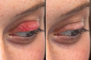 Conservative management is the initial strategy for a chalazion. This includes warm compresses on the eyelid for 15 minutes 2 to 4 times a day. Eyelid massage and possibly also use of baby shampoo on the eyelids can be effective. Most chalazia resolve within a month with these conservative measures. If the symptoms persist beyond a month, examination by an ophthalmologist is recommended. There have been reports of a change in the position of the lesion with conservative management. If this happens, referral to an ophthalmologist for surgical treatment is suggested. There is potential for larger central lesions to cause complications, so earlier referral for surgical management should be considered in these cases too. Antibiotics are not routinely required since it is an inflammatory condition. However, there may be cases in which an associated infectious etiology is suspected. If an infection is considered, tetracyclines are the preferred antibiotic. Doxycycline 100 mg orally twice a day for 10 days or minocycline 50 mg orally for 10 days would be reasonable options. In patients who are unable to take tetracycline, metronidazole is the preferred alternative. If there is no evidence of infection, steroids can be used by injection into the lesion area. An injection of 0.2 to 2 ml of a triamcinolone 40 mg/ml solution would be a typical choice. Larger lesions may require a repeat injection within 2 to 7 days. Persistent lesions require surgical intervention. Smaller lesions can be treated by surgical excision. Larger lesions require more extensive excision. A recurrent chalazion should be biopsied to rule out sebaceous cell carcinoma. An effective treatment that can prevent the need for surgery is Intense Pulsed Light treatment combined with gland expression.
Differential diagnosis
Although less common than a chalazion, tumors should be considered, especially in a recurrent chalazion among the elderly. Sebaceous cell carcinoma and squamous cell carcinoma should be ruled out by biopsy if there is clinical concern. Infectious etiologies such as blepharitis, dacryocystitis, herpes zoster, herpes simplex, molluscum contagiosum, leishmaniasis and cellulitis should be considered and treated. Benign lesions such as papillomas, hordeolum, juvenile xanthogranuloma and xanthelasma should be considered if the appearance is not typical of a chalazion.
Prognosis of a chalazion
The prognosis is excellent for patients with a chalazion. There is often a good response to conservative treatment (warm compresses and eyelid hygiene).
Complications
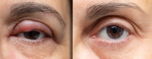 An untreated chalazion can lead to cellulitis, which can lead to deformation of the eyelid shape with progression. A large central chalazion can cause vision disturbances due to the effects of direct contact with the cornea. Involvement of the upper eyelid increases astigmatism and corneal deformation, especially in the peripheral cornea. This risk increases significantly in situations where the chalazion is larger than 5 mm. Therefore, excision of these lesions should be considered.
Consultations with a specialist
As explained, a chalazion that becomes complicated, large or unresponsive should be evaluated by an ophthalmologist.
Prevention of a chalazion
Warm compresses, eyelid hygiene, regular gland expression by a specialist physician and Intense Pulsed Light treatment in order to control the chronic eyelid inflammation (blepharitis) that is usually the number-one cause of a chalazion are critical elements for the prevention of a chalazion.
Have a question about your eyes? Want a specialist opinion?