Pterygium – Treatment

When we talk about the growth of a pterygium, its treatment takes on added importance. A pterygium is scar tissue on the surface of the conjunctiva that grows over the front part of the cornea. A pterygium usually grows slowly and over many years, and is more common in people who have been exposed to a lot of sunlight during their lives.
Is a Pterygium Always Symptomatic?
A mild pterygium can in many cases be asymptomatic or cause redness of the eye, discomfort in the eye such as a sensation of dryness or roughness, and even dissatisfaction with the aesthetic appearance of the eye.
In extreme cases, a pterygium can grow over the central part of the cornea and cause blurred vision or scarring that may limit the movement of the eye and cause double vision.
Pterygium Treatment – Is It Necessary?
When a patient is diagnosed with a pterygium, the first thing to decide is whether any treatment is actually needed. In many cases these are only mild complaints of discomfort, and often the best approach is monitoring or medical treatment. Lubrication or topical treatment with non-steroidal anti-inflammatory agents may help ease inflammation of the eye and reduce the unaesthetic appearance. Protecting the face and eyes from excessive exposure to UV radiation may also help prevent development of the condition. In principle, only a small percentage of pterygium cases require surgical intervention; however, when the condition has developed to the point of blocking the visual axis, or irregular astigmatism caused by the pterygium, removal of the pterygium by surgery is required. Surgery is also performed in certain cases due to chronic eye irritation or aesthetic considerations.
Surgical Pterygium Treatment
The main challenge to successful surgical treatment of a pterygium is recurrence, which manifests as fibrotic growth beyond the limbus onto the cornea. Many surgical techniques are used, although none of them is universally accepted because of varying recurrence rates.
The surgery is performed under local anesthesia, in which the anesthetic is injected into the soft fatty tissue around the eyeball and not only numbs the eye but also reduces vision and stops the movement of the eye.
First, an incision is made at the limbus where the pterygium begins to invade over the cornea, and it is peeled off the surface of the cornea. Once the pterygium has been removed, the cornea can be smoothed, and once the cornea has been repaired, attention turns to treating the sclera and conjunctiva. Now the Tenon's capsule in which the pterygium was located is removed. When the scar tissue has been removed from the cornea, a decision must be made about what to do with the space remaining after the removal of the scar tissue. One of the following options can be chosen.
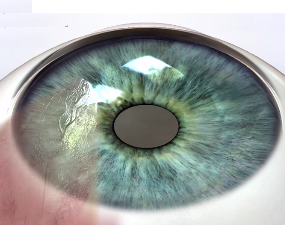
Option 1: Do nothing. The area can be left exposed and will undergo epithelialization on its own. This option carries the highest risk of pterygium recurrence, and it also causes the patient the most discomfort.
Option 2: Cover the exposed area with a biological material; an amniotic membrane is usually placed over the exposed sclera. Biological glue can be used to help graft the amniotic membrane. This method is very effective, comfortable for patients, and assists in tissue healing. It also reduces the recurrence rate.
Option 3: Autograft technique. The procedure involves obtaining a graft of healthy conjunctiva from another area of the eye and suturing the graft over the exposed sclera after excision of the pterygium. With this method, low recurrence rates have been achieved.
Mitomycin C inhibits the proliferation of fibroblasts and can be used in two ways: direct application to the exposed sclera during surgery, or treatment with eye drops after surgery. The danger with this treatment is that too high a dose can be toxic.
Recurrence Rates After Pterygium Treatment
The high recurrence rates associated with the surgery continue to be a problem, and therefore complementary medical treatments have been incorporated into the procedure and have shown a marked decrease in recurrence rates; however, they are not free of complications themselves.
Beta radiation has also been used to prevent recurrence, since it inhibits mitosis in the rapidly dividing cells of the pterygium. However, the negative effects of radiation have led physicians to recommend against its use.
Complications of Pterygium Treatment
Complications during surgery: since the surgery involves only the outer layers of the eye, pterygium surgery is relatively safe compared to many types of eye surgery.
One of the eye muscles is located partly beneath the surgical site. It is possible, though unlikely, that this muscle will be injured during surgery, which would cause double vision and require additional surgery. The possibility of this complication is of concern mainly when removing a pterygium that has regrown after previous surgery, when there is much more scarring and it is difficult to identify the muscle.
Very rarely, the internal structures of the eye may be injured and cause vision loss. This possibility is of concern when operating on an eye that has already undergone surgery or when the eye has been affected by a previous injury or another eye disease.
Complications after surgery: recurrence is the most serious complication after pterygium removal surgery. Additional complications that may be encountered after surgery include scleral melt due to the use of Mitomycin C; fibrosis, especially around the extraocular muscle in the surgical area; infection, which is rare but possible; and discomfort on the surface of the eye, which can last weeks or even months.
Steroids usually ease the sensation of discomfort, and the use of lubricant drops is also recommended.
Have a question about your eyes? Want a specialist opinion?