Dry eyelids (dermatitis of the eyelids)
Introduction to dry eyelids
Dry eyelids are usually a sign of inflammation of the skin in the area of the eyelids (eyelid dermatitis/eczema), also known as periocular dermatitis (around the eyes) or periorbital (around the orbit). Beyond a sensation of dry eyelids, it is expressed in a scaly (scales) and erythematous (redness) eruption of the upper and/or lower eyelids. Patients often report symptoms of itching, burning and stinging. There may be swelling. Dermatitis of the eyelids (dry eyelids) may be caused by contact with irritants or allergens, or it can be an expression of an underlying skin disease, such as atopic dermatitis or seborrheic dermatitis. Periorbital dermatitis may also be an expression of rosacea (ocular rosacea) or perioral dermatitis.
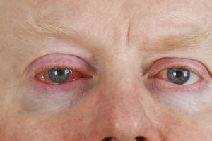
In this article we will discuss the clinical manifestations, differential diagnosis, evaluation and treatment of the most common types of eyelid dermatitis (dry eyelids). Blepharitis, chronic inflammation of the eyelid margin associated with eye irritation, is discussed in the blepharitis article. Further information about redness around the cheeks and eyelids is discussed in the rosacea article.
Epidemiology of dry eyelids
Eyelid dermatitis is usually seen in adults and adolescents and is less common in children, unless associated with atopic dermatitis. The exact prevalence of eyelid dermatitis in the general population is not known, but it is very common. Men and women alike can show signs of dry eyelids, but the prevalence of dry eyelids is higher in women than in men in most studies that have examined the subject of eyelid skin inflammation.
Allergic contact dermatitis is the most common type of eyelid dermatitis that causes dry eyelids, constituting about 50% of cases, followed by irritant contact dermatitis and atopic dermatitis.
Aetiology of dry eyelids
The cause of skin inflammation in the eyelids is usually multifactorial. It may result from contact with irritants or allergens, or it can be an expression of an underlying skin disease, such as atopic dermatitis or seborrheic dermatitis.
Contact dermatitis – the eyelids are a sensitive site because of the thinness of the skin and the increased penetration potential of allergens and irritants. Often, the eyelids may be the only site affected by such inflammation. For example, dry eyelids may be the only expression of a reaction to hair-care products. Nail products and other allergens may be transferred from the hand to the eyelid area and may cause isolated skin inflammation in the absence of dermatitis in the hand area. The classic clinical signs are dryness and itching of the eyelids.
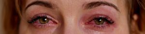
Allergic contact dermatitis – allergens found to cause dry eyelids and dermatitis include metals (for example, nickel, gold), fragrances, preservatives and topical antibiotics.
Topical ocular preparations may cause dermatitis as a result of contact and dryness in the eyelids. In a study carried out on 4779 patients in Europe with dry eyelids, two distinct populations with allergic eyelid dermatitis were identified: younger patients with a history of atopic dermatitis and frequent use of cosmetics, and older patients with an allergy to topical ocular preparations. Allergens found in topical ocular preparations included phenylmercuric acetate, topical antibiotics (including gentamicin and neomycin) and phenylephrine.
Airborne contact dermatitis – airborne contact dermatitis of the eyelids is caused by exposure to airborne antigen or irritant particles. These include plant antigens, tree allergens, plastic, rubber, glue, metal, industrial and agricultural dust, pesticides and medications.
Protein contact dermatitis – protein contact dermatitis occurs mediated by immunoglobulin E (IgE) due to sensitivity to proteins from plants or animals. These include food, pollen, animal hair and latex.
Irritant contact dermatitis – irritants known to cause or worsen eyelid dermatitis and cause dry eyelids include soaps, preservatives and fragrances. Use of anti-ageing products on the face may cause skin inflammation of the eyelids even if the product is not used directly on the eyelids.
Atopic dermatitis – eyelid dermatitis may appear in patients with a history of atopic dermatitis and/or other atopic diseases, including asthma and seasonal allergies. Irritants often play a role in the development of eyelid dermatitis in patients with atopic disease. In addition, these patients may develop dermatitis as a result of contact and even dermatitis secondary to exposure to proteins as mentioned earlier. All of these may lead to dryness and itching of the eyelids and irritation.
Seborrheic dermatitis – seborrheic dermatitis is a chronic, recurrent form of dermatitis, which has a tendency for areas with folds such as the nose, eyelids, ears, scalp, chest and other sites. Less commonly, it may involve the skin of the eyelid and/or the eyelid margin (seborrheic blepharitis). In a series of 447 patients with eyelid dermatitis or dry eyelids, 6 percent were diagnosed as suffering from seborrheic skin inflammation.
Signs and clinical manifestations of dry eyelids
Dry eyelids or eyelid dermatitis usually presents as an erythematous, scaly rash, with irritation of the upper and/or lower eyelids. It may occur separately or be associated with skin inflammation of other body sites. Dermatitis is in most cases bilateral, but may be unilateral and affect the upper or lower eyelids, or both. The clinical findings may vary according to the aetiology.
Patients with contact eyelid dermatitis present with itching, scales, burning and pain in one or both eyelids. Itching is often a prominent symptom of allergic contact dermatitis, and there may be features of worsening as a result of chronic rubbing and scratching. In a series of 264 patients with eyelid dermatitis, common symptoms included erythema, itching, oedema and scaling. Beyond dry eyelids they may appear cracked, and a crust may appear on the lashes. About 25% of patients suffering from contact eyelid dermatitis also suffer from inflammation of the conjunctiva. Dermatitis often appears in other places on the body, including the face, the neck and the periauricular skin.
Atopic eyelid dermatitis often develops in adolescence and in young adults but may also occur in older people. Occasionally, it can be the only expression of atopic dermatitis. The upper eyelids may appear scaly and cracked. Common signs are "allergic shiners" (symmetrical, dark circles under the lower eyelid) and Dennie-Morgan lines (additional skin folds under the lower eyelid).
However, it is sometimes difficult to distinguish between atopic eyelid dermatitis and skin inflammation secondary to contact. In addition, patients with atopic dermatitis may also develop skin inflammation secondary to contact.
Seborrheic skin inflammation on the eyelids may appear more scaly and waxy than dermatitis secondary to contact or atopic dermatitis; itching is less common than other symptoms, such as scaling (dry eyelids) or burning.
Clinical course of dry eyelids
The clinical course often varies with the aetiology. Eyelid skin inflammation may come and go intermittently and sometimes presents a seasonal variation. For example, eyelid dermatitis associated with atopic dermatitis may be chronic and flare up with the seasons of the year and exposure to environmental allergens. Patients may notice that airborne irritants, including exposure to plants, pollen, dust and dandruff, and that these may cause or worsen the disease. Eyelid dermatitis may also flare up with hay fever, allergic rhinitis or conjunctivitis, associated perhaps with chronic watering or mechanical rubbing. Occupational eyelid dermatitis may worsen with exposure to certain conditions in the workplace and improve with time off work.
Diagnosis of dry eyelid
Clinical – the diagnosis of eyelid dermatitis is made in most cases clinically, based on the characteristic appearance of the eruption, accompanying symptoms and clinical history.
A careful clinical history may provide clues to the aetiology. As an example, the presence of bilateral eyelid dermatitis, prominent itching, involvement of other sites on the face, lack of seasonal variation of the symptoms and a history of temporary or poor improvement with standard treatment indicate a diagnosis of allergic contact dermatitis.
Atopic skin inflammation is suspected in adolescents and young people with a history of atopic dermatitis and/or other atopic diseases, including asthma and seasonal allergies. The presence of dermatitis in the area of the joints supports the diagnosis. However, exposure to irritants or a component of contact dermatitis may play a role in the development of eyelid dermatitis in patients with atopic disease.
Patch testing – atopic eyelid dermatitis and contact eyelid dermatitis can sometimes be clinically indistinguishable. Every patient with eyelid dermatitis requiring ongoing treatment beyond four to eight weeks should be considered for patch testing. If the patch tests are negative or positive reactions are not relevant to the current skin inflammation, the diagnosis of irritant contact dermatitis should be considered.
Skin biopsy – a skin biopsy is usually not helpful for the diagnosis of skin inflammation in the eyelids. Histopathological examination will show non-specific changes common to all types of eczematous dermatitis and will therefore not help identify the specific cause of skin inflammation in the eyelids. However, a skin biopsy can be carried out if the diagnosis is uncertain and especially to rule out connective tissue diseases and dermatomyositis.
Associated eye diseases
Most patients with eyelid skin inflammation have no additional associated eye disease. However, patients with atopic dermatitis have an increased risk of accompanying eye diseases such as blepharitis (inflammation of the eyelids), keratitis (inflammation of the cornea), conjunctivitis and keratoconus.
Differential diagnosis of dry eyelids
Further causes of dryness in the eyes include: psoriasis, rosacea and dermatomyositis.
Treatment of dry eyes and dermatitis of the eyelids
The treatment of eyelid skin inflammation includes ongoing avoidance of exposure to irritants and allergens for patients with contact dermatitis and the use of topical anti-inflammatory agents, including topical steroids and topical calcineurin inhibitors (such as tacrolimus).
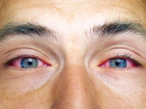
Skin care – conservative initial management of eyelid dermatitis includes gentle care of the skin and avoidance of perfume and other irritants in personal, hair and facial skin-care products. Fragrance-free emollients, such as petrolatum, can be applied directly to the eyelids.
Avoidance of irritants and allergens – for patients with a confirmed diagnosis of contact or allergic eyelid dermatitis, ongoing avoidance of irritants and allergens is the mainstay of treatment. Petrolatum or other ointment-based emollients, free of fragrance and other common allergens, can be used. The use of perfume and hair sprays should be avoided.
Topical steroids – topical steroids and not calcineurin inhibitors can be offered as first-line treatment for eyelid skin inflammation. Because the eyelids demonstrate high absorption, only low-potency steroids are safe for long-term use on the eyelids. For prolonged treatment it is preferable to move to a topical calcineurin inhibitor. Prolonged use of topical steroids in the eyelid area may cause several side effects including thinning and atrophy. Long-term use of topical steroids on the eyelids can also lead to the development of skin inflammation and a rosacea-like eruption. Prolonged use of steroids in the eye area can also cause an increase in intraocular pressure and the development of glaucoma.
Topical calcineurin inhibitors – topical calcineurin inhibitors (tacrolimus and pimecrolimus) can be used as an alternative to topical steroids for the treatment of skin inflammation in the eyelids in patients requiring prolonged treatment (beyond four weeks). Calcineurin inhibitors are applied twice a day. Initially there may be a burning sensation with use of the preparation but this tends to improve over time.
Have a question about your eyes? Want a specialist opinion?