What is corneal topography?
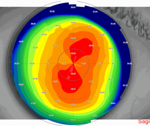 Corneal topography makes it possible to assess, at very high resolution, the integrity of the cornea. It is possible to assess the shape of the cornea, its curvature and its thickness profile with the help of several technologies. Topography mapping is based on a Placido disk (Placido disk) and provides mainly information about the curvature and the optical power of the cornea in different areas. Tomography mapping provides information about the elevation and thickness in different areas of the cornea and is based on Scheimpflug technology (Scheimpflug) or scanning slit (scanning slit). Today these systems can also incorporate the functionality of measuring pupil size, pupil position, regular and irregular astigmatism, automatic detection of keratoconus, keratometry, corneal asphericity, aberrometry based on wavefront technology (wavefront aberrometry) and topographic information.
Keratoconus
August 19, 2023Keratoconus (Keratoconus). Keratoconus of the cornea of the eye (keratoconus) is a disease that appears in 0.05% of the population. In this disease the central or paracentral (paracentral) part of the cornea undergoes thinning and protrusion so that the cornea takes on the shape of a cone (and from there comes
Corneal cross-linking – cross linking (Corneal Cross Linking)
August 17, 2023Corneal cross-linking (cross linking). Corneal cross-linking (cross linking) (Corneal Cross Linking) brought a revolution in our ability to treat keratoconus and ectasia of any cause and to prevent the progression of this common disease. Keratoconus
Interpretation of corneal mapping
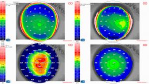
October 1, 2021A corneal mapping interpretation service. Do you need a corneal mapping interpretation service? Looking for corneal mapping results? Send a WhatsApp message to the number 058-644-5151 with the images of your mapping together with the reason for the referral for corneal mapping, and a specialist
Topography of the cornea
Topography of the cornea provides highly detailed information about the curvature of the cornea. Assessment of the topography is performed using keratoscopic images, captured from Placido disk patterns reflected from the tear film laid on the surface of the cornea and then converted into computerized color scales. Because the image is created from the front surface of the tear film, irregularities in the composition or volume of the tears can have a great effect on the quality and results of a Placido-disk-based system. Because of this effect, reviewing the Placido image (the rings image) before interpreting the maps and numerical data afterward is extremely important. In addition, one must remember that the center point of the rings is not necessarily the patient's line of sight or the visual axis, which may lead to confusion in interpreting the topographic maps.
Corneal mapping and astigmatism
Regular astigmatism (regular astigmatism)
A normal topographic image of the cornea without astigmatism demonstrates a relatively uniform color pattern in the center with flattening in the periphery. Regular astigmatism has symmetrical and uniform steepening along a single corneal axis that is fully correctable with a cylindrical lens (glasses). A topographic image of regular astigmatism demonstrates a symmetrical "bowtie" pattern (bowtie pattern) along a single axis with a straight axis on both sides of the center. This pattern is an artifact of Placido-based topographic maps because the Placido image cannot detect the curvature at the most central point in the cornea.
Irregular astigmatism (irregular astigmatism)
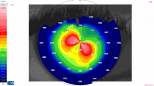
Irregular astigmatism is asymmetrical steepening of the cornea for various reasons that cannot be fully corrected by a cylindrical lens (glasses). Irregular astigmatism reduces the corrected visual acuity and may reduce contrast sensitivity (contrast sensitivity) and increase visual aberrations (visual aberrations) depending on the size of the irregularity. And hard contact lenses can improve reduced visual acuity resulting from irregular astigmatism of the cornea by bridging the irregular corneal surface and the contact lens with the tear film.
Disadvantages of topography of the cornea
The accuracy of topography of the cornea can be affected by an unstable tear film, a non-straight line between the patient's eye and the examination device, an out-of-focus image, non-measurement of the corneal periphery, less accurate after glasses-removal surgeries, less accurate in corneas with scars.
Tomography mapping of the cornea
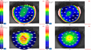
While the curvature of the cornea (and correspondingly the power) on the surface is best measured by imaging with Placido-based technology, the overall shape of the cornea, including the thickness profile, is best measured by corneal mapping of the tomography type. There are various imaging systems that photograph multiple slit images and reconstruct them into a corneal-shape profile, including anterior and posterior corneal elevation data. These include scanning slit technology (scanning slit), Scheimpflug-based imaging systems (Scheimpflug) and Anterior Segment Optical Coherence Tomography (Anterior Segment Optical Coherence Tomography). In general, each device calculates the best fit sphere (best fit sphere) for each map separately. For this reason, comparing the elevation maps between devices in the same patient is inaccurate because they usually have different best fit spheres. Elevation-based tomography is especially useful in refractive surgeries (glasses removal) for describing the shapes of the anterior and posterior surfaces of the cornea. With such information it is possible to determine changes in the shape of the corneal structure more accurately over time or before and after treatment.
Corneal mapping and diagnosis of keratoconus (corneal mapping keratoconus)
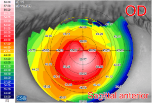
Topography maps of the cornea make it possible to identify keratoconus, even in the early stages, and also to identify who is at risk of developing ectasia after refractive surgery (laser glasses removal). A regular astigmatic profile has a bowtie pattern. Keratoconus can be identified by a central cone or inferotemporal elevation. A non-straight or symmetrical astigmatic axis with inferior steepening also indicates keratoconus. The ratio between the lower and upper keratometry values can indicate keratoconus. A difference greater than 1.4 diopters between the two and a central curvature greater than 47.2 diopters can indicate ectatic disorders of the cornea, but there is some overlap between normal and abnormal eyes. The disease called Pellucid Marginal Degeneration has a characteristic "crab claw" configuration with horizontal astigmatism (against the rule). However, this pattern is not reserved for PMD, because it can also be seen in keratoconus (inferior keratoconus). Topography of the cornea can help in accurate monitoring of the progression of these ectatic corneal diseases and identify patients who would benefit from corneal cross-linking treatment (cross-linking). Previous glasses-removal surgery will demonstrate central flattening in myopia corrections and central steepening in hyperopic corrections.
Prof. Michael Mimouni is a specialist in corneal mapping keratoconus.
Have a question about your eyes? Want a specialist opinion?