The cornea plays a vital role in our ability to see the world around us. It is a transparent, dome-shaped tissue located at the front of the eye that acts as a protective barrier and focuses light on the retina. Here we will delve into the anatomy of the cornea, common diseases of the eyes' corneas, the significance of corneal topography tests, the emergence of artificial corneas, the life-changing procedure of corneal transplant surgery, and the importance of corneal donation.
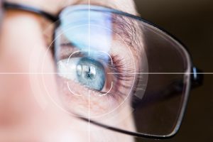
Anatomy of the Cornea:
The cornea is a clear, avascular structure that covers the front part of the eye. It is composed of five separate layers. The cornea works in conjunction with the lens to refract light and focus it on the retina. The outermost layer, the epithelium, acts as a protective barrier against foreign particles and bacteria. The middle layer, called the stroma, makes up most of the thickness of the cornea and provides it with strength and flexibility. Finally, the innermost layer, the endothelium, maintains the transparency of the cornea by regulating its moisture.
Diseases of the Cornea:
Various conditions can affect the corneas and lead to vision impairment. Some common corneal diseases include:
- Corneal dystrophy: hereditary disorders that cause progressive changes in the structure of the cornea, leading to vision loss.
- Keratitis: inflammation of the cornea, usually caused by infections, contact lens use, or injuries.
- Corneal abrasions: scratches on the surface of the corneas due to trauma or foreign objects.
- Corneal ulcers: open sores in the cornea, usually caused by infections or severe dryness in the eyes.
- Keratoconus: a condition in which the cornea gradually thins and bulges, resulting in distorted vision.
Corneal Mapping – Corneal Topography:
Corneal topography is a non-invasive diagnostic test that provides detailed information about the shape, curvature, and thickness of the cornea. It involves projecting illuminated patterns onto the cornea and capturing their reflection to create a topographic map. This test is used to detect irregularities in the cornea, determine the suitability of contact lenses, assess corneal diseases, and plan various eye surgeries such as LASIK.
Artificial Cornea:
In cases where a damaged cornea cannot be restored using conventional treatments, artificial corneas, also known as keratoprostheses, offer a treatment option. These devices are designed to replace the damaged cornea and restore vision that has been impaired. Artificial corneas are usually of a biocompatible type and can be implanted through a surgical procedure. Artificial corneas are used mainly when a traditional corneal transplant surgery is not a practical option.
Corneal Transplant Surgery:
Corneal transplantation is a surgical procedure that involves replacing a diseased or damaged cornea with a healthy donor cornea. It is a highly successful procedure and can restore vision in people with diseases or injuries of the cornea. There are different types of corneal transplants, depending on the extent of the damage to the cornea.
Corneal Donation:
Corneal donation is an act that can change lives. Donated corneas are received from people who have passed away. The cornea is especially suitable for transplantation because it does not require blood-type matching.
In Summary:
The cornea, with its complex structure and vital role in vision, deserves the attention and care required. From diagnosing corneal conditions using topography tests to restoring vision through corneal transplant surgery or the use of an artificial cornea, advances in ophthalmology continue to expand the treatment options.
Have a question about your eyes? Want a specialist opinion?