Corneal topography interpretation service
Do you need a corneal topography interpretation service? Looking for corneal topography results? Send a WhatsApp message to the number 058-644-5151 with the images of your topography together with the reason for the referral for corneal topography, and a cornea specialist (an ophthalmologist who is an expert in the field) will go over your topographies and perform the interpretation.
Four main reasons for referral to corneal topography, together with examples and interpretations
Keratoconus
Keratoconus is a disease in which there is thinning and steepening of the cornea that ultimately leads to impaired vision. On your left is an example of a corneal topography of a patient with keratoconus. Corneal topography interpretation of keratoconus: on the thickness map (upper left) one can see that there is thinning of the cornea (yellow) in an area that is slightly inferior and temporal (toward the temple) to the center of the cornea.

Upper right is the curvature map of the cornea, and here one can see that in a corresponding area there is high curvature of the cornea (red and white colors) indicating a "bulge" in the cornea in this area. In addition, there is anterior elevation (lower-left map) and posterior elevation (lower-right map) in corresponding areas in these maps. In summary: thinning of the cornea, high curvature, anterior and posterior elevation, all in the same area of the cornea (inferior and temporal). Classic signs of keratoconus.
Pellucid Marginal Degeneration
Pellucid Marginal Degeneration, also known by the abbreviation PMD, is very similar to keratoconus (and some even claim that it is the same spectrum).
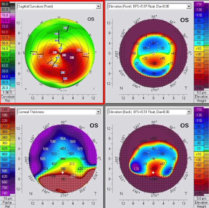
It usually involves older patients than patients with keratoconus, and the hallmark of the disease is marked thinning in the lower part of the cornea, with the area of thinning located below the area of highest curvature. Corneal topography interpretation of PMD: on the thickness map (lower left) one can see that there is very impressive thinning in the lower part of the cornea.
Upper left in the curvature map there is a classic sign of an against-the-rule "crab claw" or butterfly configuration. Sometimes it is very difficult to distinguish between inferior peripheral keratoconus and PMD, and therefore some consider them to be different variants of the same disease.
A cylinder value that has recently increased
Corneal topography interpretation of a regular cylinder
Patients are often referred for corneal topography because there is an increase in the cylinder.
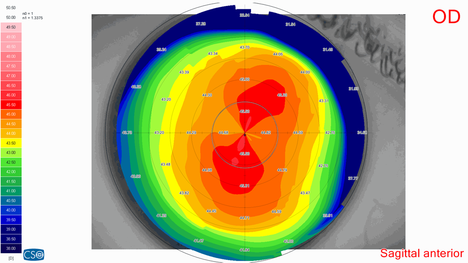
It is important to distinguish between two types of cylinder. Normal corneal topography interpretation: a regular cylinder (regular astigmatism) in which there is steepening in one axis and which is fully correctable with glasses. In such a cylinder we expect to see a bow-tie pattern that is straight and symmetrical, as can be seen in the image on the left. Such a state of symmetrical cylinder usually does not require surgical intervention, and one can simply follow up with repeat topographies over time to make sure it is stable.
Corneal topography interpretation of an irregular cylinder
In contrast, there are situations in which the cylinder is not entirely regular and has an irregular component. In such a state, there is already no very good vision with glasses alone, and patients will report that they see better with rigid contact lenses. Irregular corneal topography interpretation:
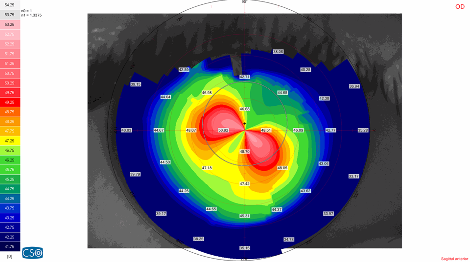
As can be seen in the topography on the left, the two sides of the bow-tie (the red balloons) do not sit on the same axis. This can be an early sign of various diseases such as keratoconus or PMD, but it can also remain stable in this state. One must proceed with extra caution before performing laser vision-correction surgery on such an eye, and it is advisable to follow up on such an eye periodically to make sure it is stable.
Corneal topography interpretation prior to laser vision-correction surgery
Every patient who is about to undergo laser vision-correction surgery must undergo corneal topography, and this is for several reasons.
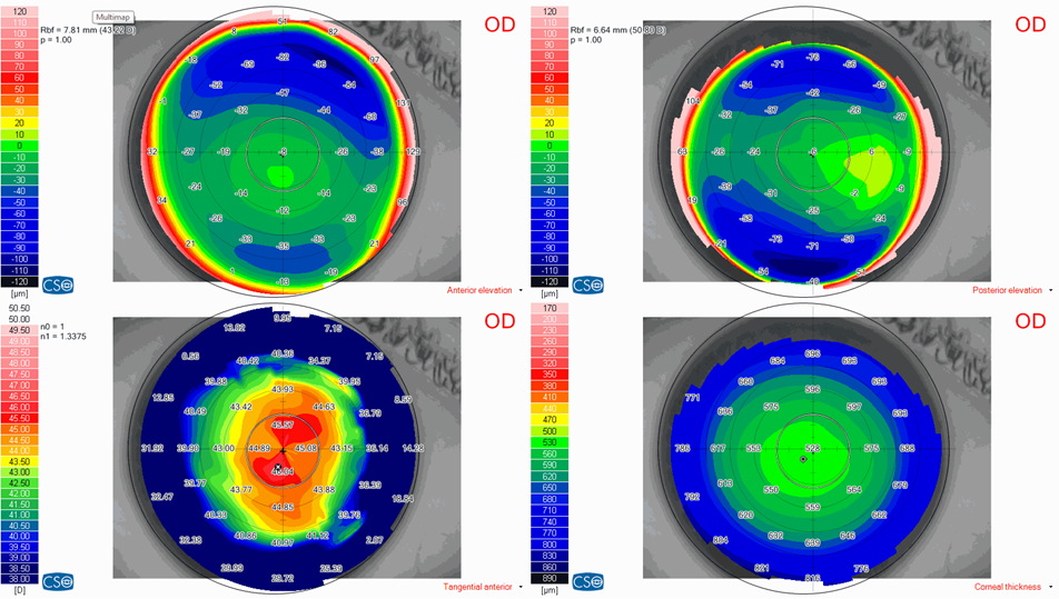
The first reason is to rule out a corneal disease that constitutes a contraindication to performing laser vision-correction surgery. In addition, the corneal topography will allow the surgeon to choose the most suitable surgery for the patient (PRK, LASIK or SMILE). Corneal topography interpretation before laser vision correction: here one can see that there is a normal corneal topography with normal thickness (around 528 microns) in the lower-right map. In addition there is no anterior or posterior elevation of the topography, and there is central curvature that is regular and of normal strength. Such a patient will most likely be able to undergo laser vision-correction surgery, and assuming they do not have too high a number, they will be able to undergo both LASIK and PRK.
Have a question about your eyes? Want a specialist opinion?