Background for an artificial corneal transplant
An artificial corneal transplant is based on the use of a corneal graft that is made, at least in part, of synthetic raw material, and is intended to provide an optical, structural and functional answer for patients who suffer from blindness caused by disease in the cornea of the eye. There are about 4.9 million people with bilateral blindness secondary to corneal disease worldwide, constituting 12% of total global blindness. The common causes are pathologies of the anterior cornea such as trachoma, infectious keratitis, eye trauma and chemical injuries - all of these are more common in developing countries. A primary corneal transplant (lamellar corneal transplant or full-thickness corneal transplant) has a high graft survival of 87% and 93% after one year and 72% to 73% after 5 years in non-complex eyes. Transplant survival decreases in eyes that have undergone repeat transplants and in complex eyes, despite advances in transplantation techniques and more selective tissue transplantation. It is possible that these eyes with a high risk of graft failure will achieve a better result with an artificial corneal transplant.
Risk factors and indications for an artificial corneal transplant
High risk factors include recurrent and chronic inflammation of the external surface of the eye, glaucoma and eyes with numerous blood vessels in the cornea. Worldwide, the availability of raw material (a cornea from a donor) can be limited due to the supply of donors and the need for eye-bank facilities. An artificial corneal transplant can be considered for complex corneal diseases such as multiple transplant failures or severe inflammatory disease of the eye surface (after a burn, Stevens-Johnson, etc.).
Corneas and devices used for an artificial corneal transplant
Many devices for an artificial corneal transplant (keratoprosthesis; KPro) have been proposed. Pellier de Quengsy first described the initial idea in 1789. They usually have a central transparent optic with rigid skirt plates between which donor corneal tissue is contributed, or a soft optic and skirt. The importance of a suitable skirt material with integration of the external eye-surface tissues was clarified from earlier models that were made of rubber, milk protein, Dacron, crystal, glass and celluloid, which led to extrusion of the graft later after the transplant. Over the years, the improvement in natural corneal-transplant methods (from a donor cornea only) caused a decline in interest and development of the artificial corneal transplant. However, the discovery of polymethyl methacrylate (PMMA) made it possible to implant a biocompatible device.
Recently, soft polymers have been used to mimic the natural cornea. Poly-2-hydroxyethyl methacrylate was used for the AlphaCor, which received FDA approval in 2003. After one year and two years, the retention rates were 80% and 62% respectively, and melting of the stroma, and loss of the graft, occurred in 27% of all cases. A similar design using polytetrafluoroethylene (PTFE; Legeais BioKPro-III) led to worse results, with 86% of the devices failing after the transplant. The focus of this article is to describe indications and management for the most common artificial corneal transplants today: Boston KPro type 1 and the Osteo-odonto-Keratoprosthesis (OOKP).
Current keratoprostheses
Boston KPro Type I
The most commonly implanted artificial corneal transplant is the Boston KPro type 1, first presented by Dohlmann in 1965 and receiving FDA approval in 1992. Its popularity began to rise from the start of the 21st century, and to date more than 19,000 such devices have been implanted. The design consists of a front plate with a central optical part, a back plate, and a donor corneal button between them. 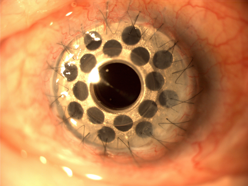 The front and optic plates are made of PMMA, and the optical power is determined by the curvature of the radius. The original design involved screwing the back plate into place. This was improved with a titanium locking ring in 2003 and a threadless stem in 2007. The back plate is available in PMMA and in titanium, both of which are well tolerated biologically. No difference was reported in the frequency of retroprosthetic membrane (RPM) formation between the two materials after 12 months.
Osteo-odonto-Keratoprosthesis (OOKP)
The OOKP uses a bone complex of a tooth-alveolar root as a keratoprosthesis skirt material for better tissue integration. Invented by Strampelli and later modified by Falcinelli, the principle of the OOKP surgery is a bypass of the diseased eye surface by using buccal mucosa and replacing the anterior-segment structures with the OOKP. The mucosal graft can tolerate dry environmental conditions and a certain level of inflammation. The good tissue integration ensures that the OOKP can be preserved for a longer time. Long-term anatomical preservation is good, with 81% preservation over 5 years reported in a group of 36 eyes, 98% preservation in 85 patients during 20 years of follow-up, and 80% in 224 eyes over 18 years.
Artificial corneal transplant – future developments
There are a large number of alternative keratoprostheses in development. Studies found that the 5-year survival both for anatomical preservation and for functional recovery were higher for the Boston type 1 KPro compared with the Aurolab keratoprosthesis. However, these were not statistically significant. Therefore the Aurolab keratoprosthesis can constitute an alternative to the Boston type 1 KPro if there are reasonable limitations or availability issues. The Lucia keratoprosthesis is a modification of the Boston KPro type 1 designed to improve the cost. The machining time was reduced by changing the locking interface between the front and back plates. Photo-etching was used instead of using a lathe, and the round holes in the back plate were replaced with radial grooves. Titanium with anodizing allowed changes in the color of the back plate to improve the appearance.
Several artificial corneal grafts are being tested for eyes with defective blinking and dry or scarred eyes. The Lux keratoprosthesis consists of a cone-shaped PMMA cylinder, a titanium sleeve and a 7.8 mm titanium back plate. A donor cornea is double-shaped centrally at 3 mm and at 7.5 mm peripherally. The PMMA cylinder is secured in the titanium sleeve and positioned through the central 3 mm opening in the donor cornea. The back plate is secured and sutured in place in the host after removing the patient's cornea with interrupted nylon sutures. A mucous-membrane graft is sutured with an opening for the optic of the PMMA cylinder. Short-term results with good preservation and functional outcomes have been reported.
Improvements in skirt materials can further improve the development of the keratoprosthesis. The OOKP is at risk of bone resorption, and a synthetic substitute with a hydrogel compound of nano-hydroxyapatite crystals (nHAp) coated with poly-lactic-co-glycolic (PLGA) microspheres is in the laboratory. A biological material based on titania-oxide graphene was implanted in vivo in a rabbit cornea without causing an immune or inflammatory reaction and could be a potential new skirt material for a keratoprosthesis.
Have a question about your eyes? Want a specialist opinion?